Cardiology ultrasound: veterinary clinical solutions
Safe, painless, and highly informative, echocardiography has become a gold standard to diagnose most heart disease cases in our companion animals with cardiac issues, allowing a clear view of the heart chambers, their contraction patterns, the heart valves and the vessels originating from the heart.

Based on clinical needs, Esaote has developed a comprehensive cardiology package and advanced solutions dedicated to veterinary practice, delivering expert diagnosis for animals of all sizes in this application.
INTERVIEWS
Testimonials
![]() VIDEO
VIDEO
The experience with ultrasound VET line
Dr. Claudio Bussadori

![]() VIDEO
VIDEO
Cardiac hemodynamic evaluation
Dr. Luca Ferasin
Dedicated VET workflow for cardiological examinations
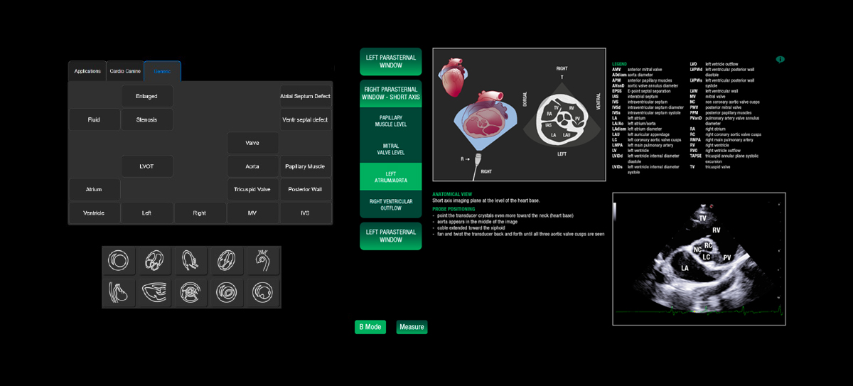
During an echocardiography, a VET-friendly and cardio-oriented interface can give an extra value to your examination.
Based on modern technologies and designed with simplicity in mind, Esaote VET Software guarantees easy workflow features, veterinary cardiological measurements whatever the current ultrasound mode used, and a dedicated report with dedicated heart’s icons.
Dedicated Cardio presets have been embedded to assist veterinarians to offer the best image settings to produce optimum visualization. With MyLibrary Equine onboard, it can support and guide you to increase the accuracy of procedures in real-time imaging.
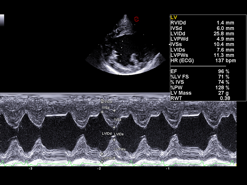
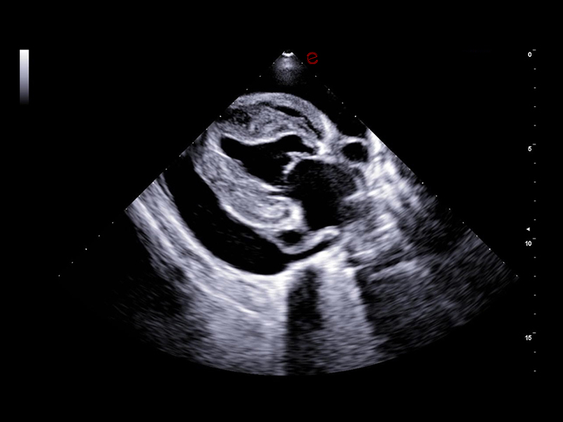
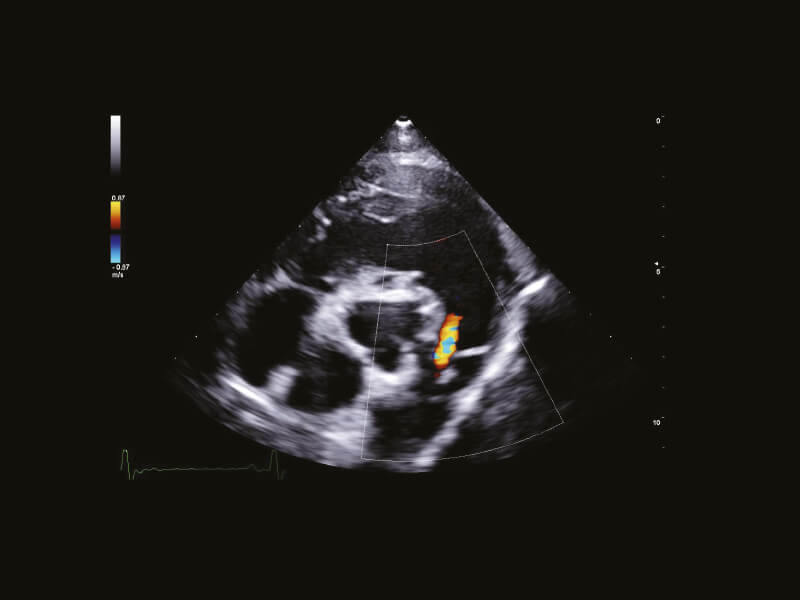
Clear Representation of Cardiac Muscles and Flows
The importance of imaging diagnostic is crucial in identifying, staging, and defining an appropriate treatment for animals of all sizes who suffer from cardiovascular diseases.
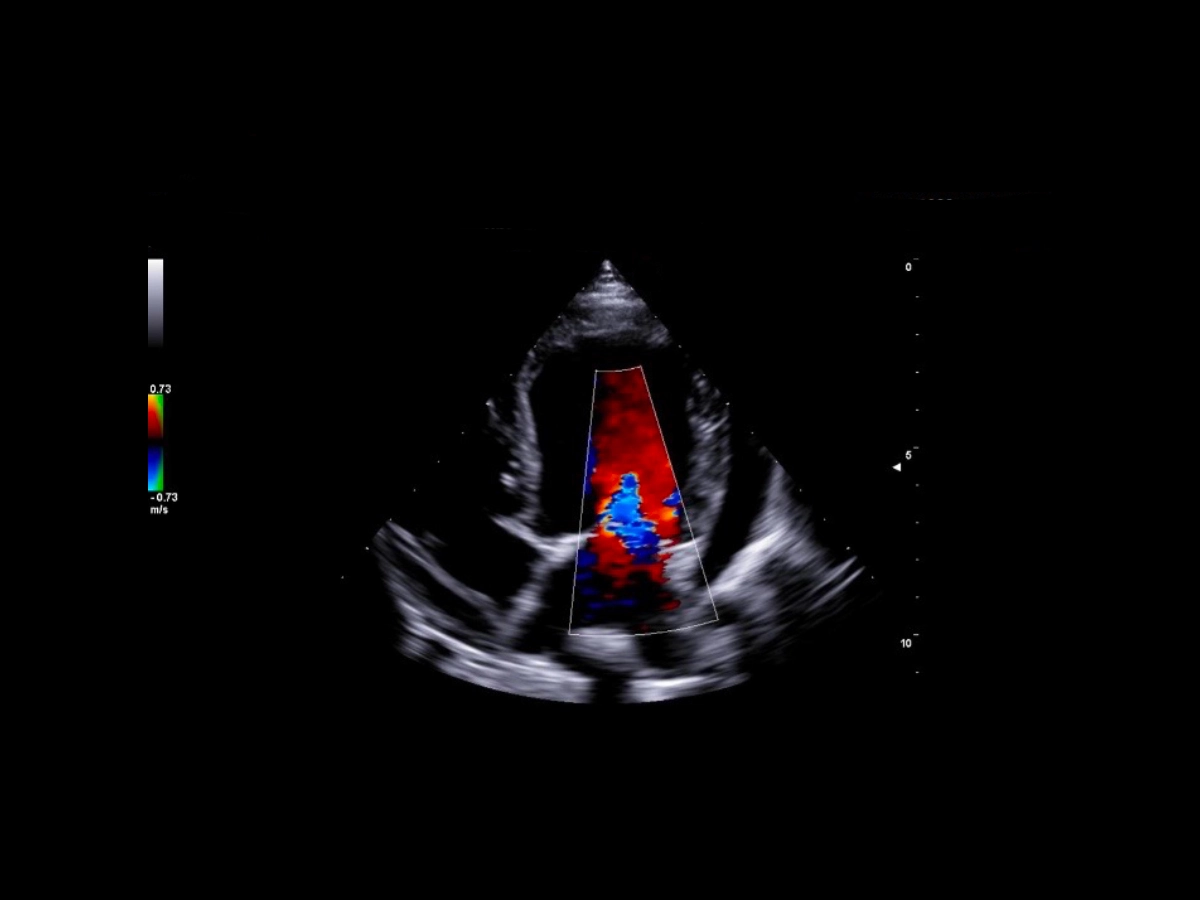
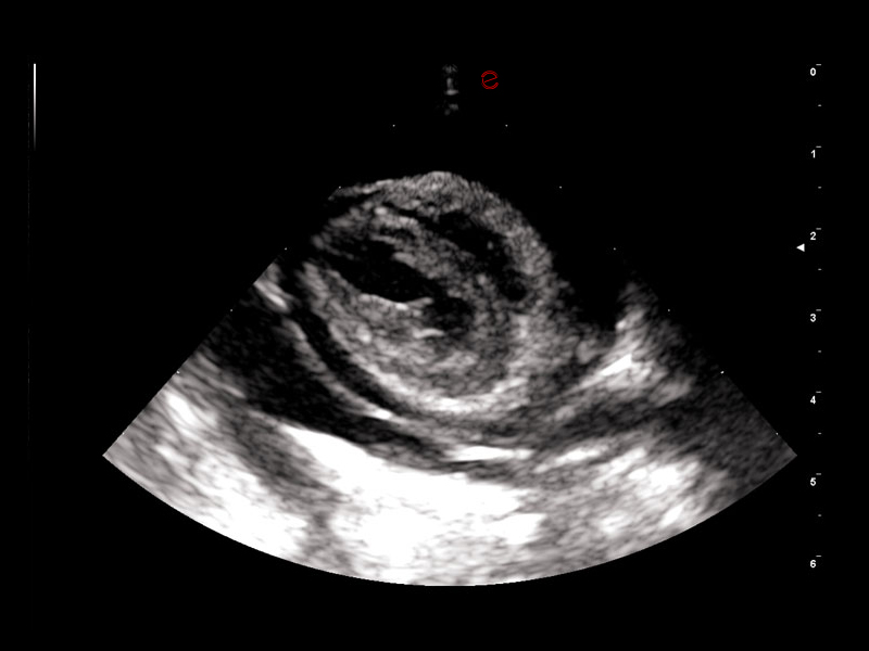
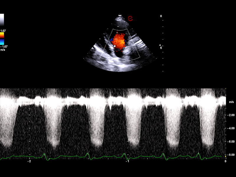
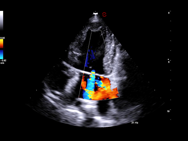
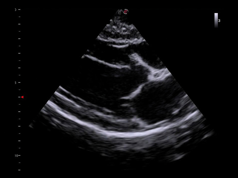
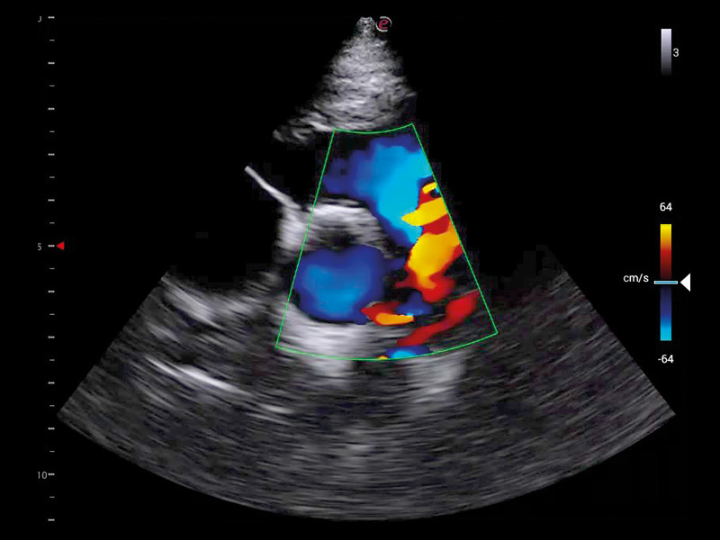
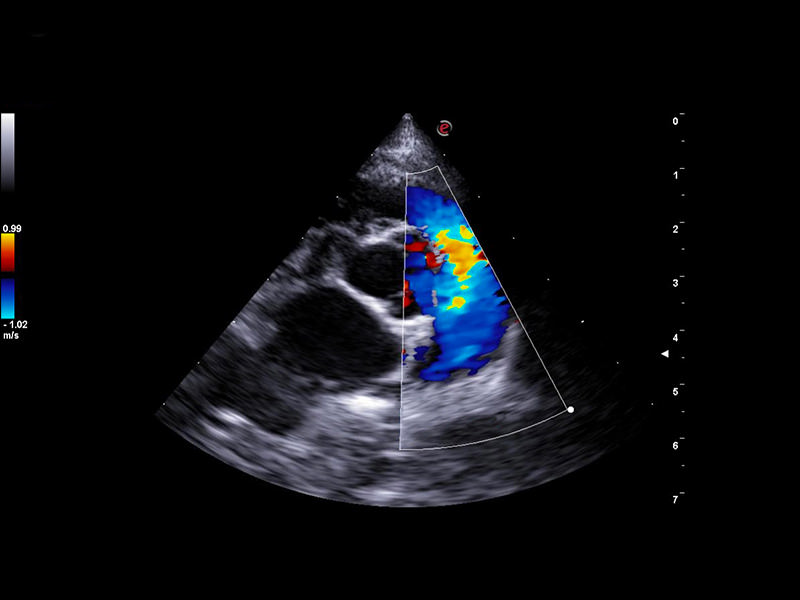
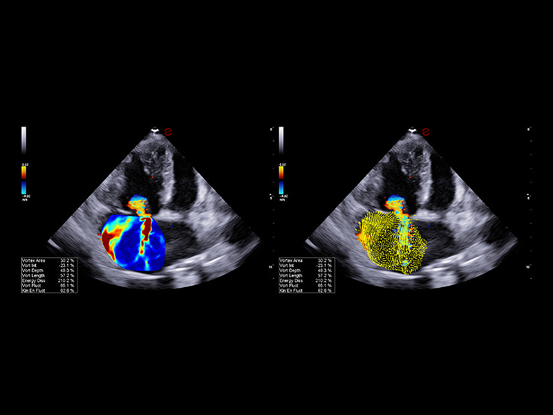
With Esaote, B-Mode and Color Doppler high performances are guaranteed to execute a precise analysis of the cardiac muscle and visualize the myocardium and valves alterations. Thanks to the state-of-the-art CFM (Esaote Color Doppler Technology) algorithm, you will obtain a realistic representation of the cardiac flows with an exceptional fluidity and filling-up, while CW (Esaote Continuous Wave Doppler Technology) and PW (Esaote Pulse Wave Doppler Technology) help identify and quantify even the smallest valve’s insufficiency.
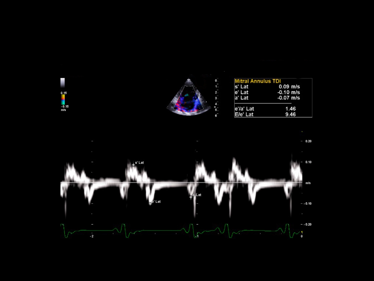
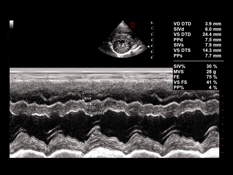
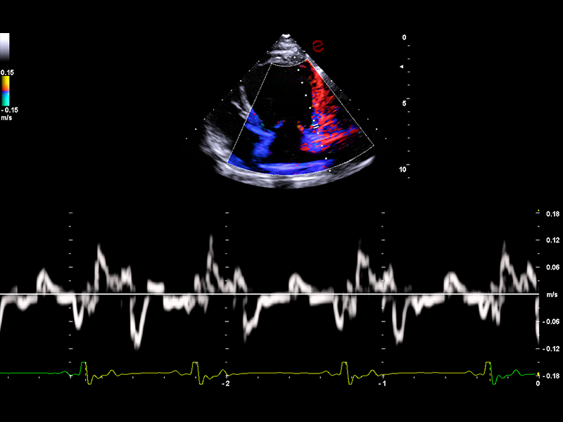
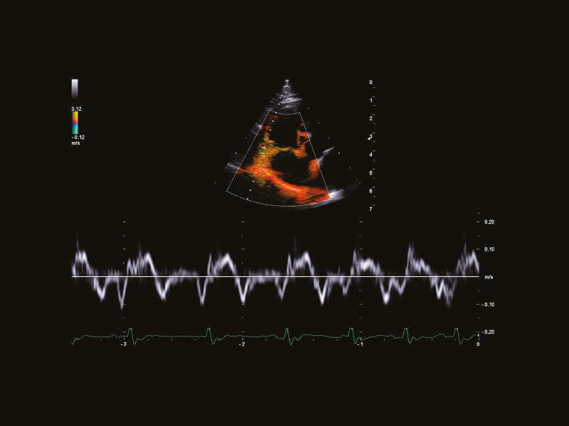
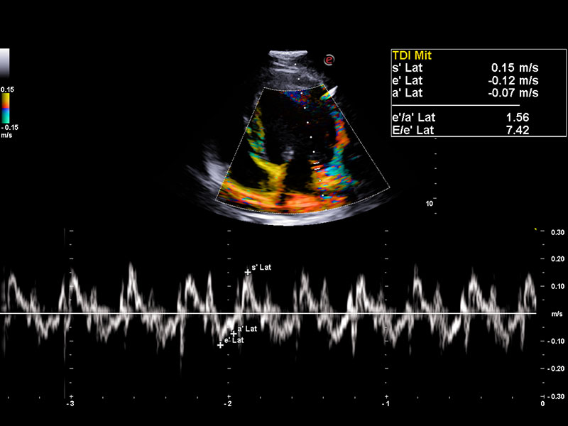
The visual estimation of wall motion might be subjective and therefore highly operator dependent, allowing only limited evaluation of radial displacement and deformation, with no possibility of assessing myocardial shortening and twisting. However, Esaote TVM (Tissue Velocity Mapping) Technology enables reliable visualization of heart wall motion, providing a comprehensive Wall Motion Analysis for evaluating both systolic and diastolic myocardial function and facilitating quantitative assessment.
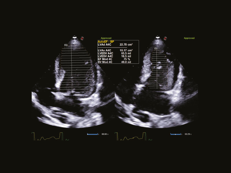
A.I.-based measurements to obtain diagnostic confidence
A standard cardiac examination includes a study of systolic, diastolic and heart valve functions. For each one, the Esaote systems can cover all the cardiology needs with specific automatization and interactive technologies.
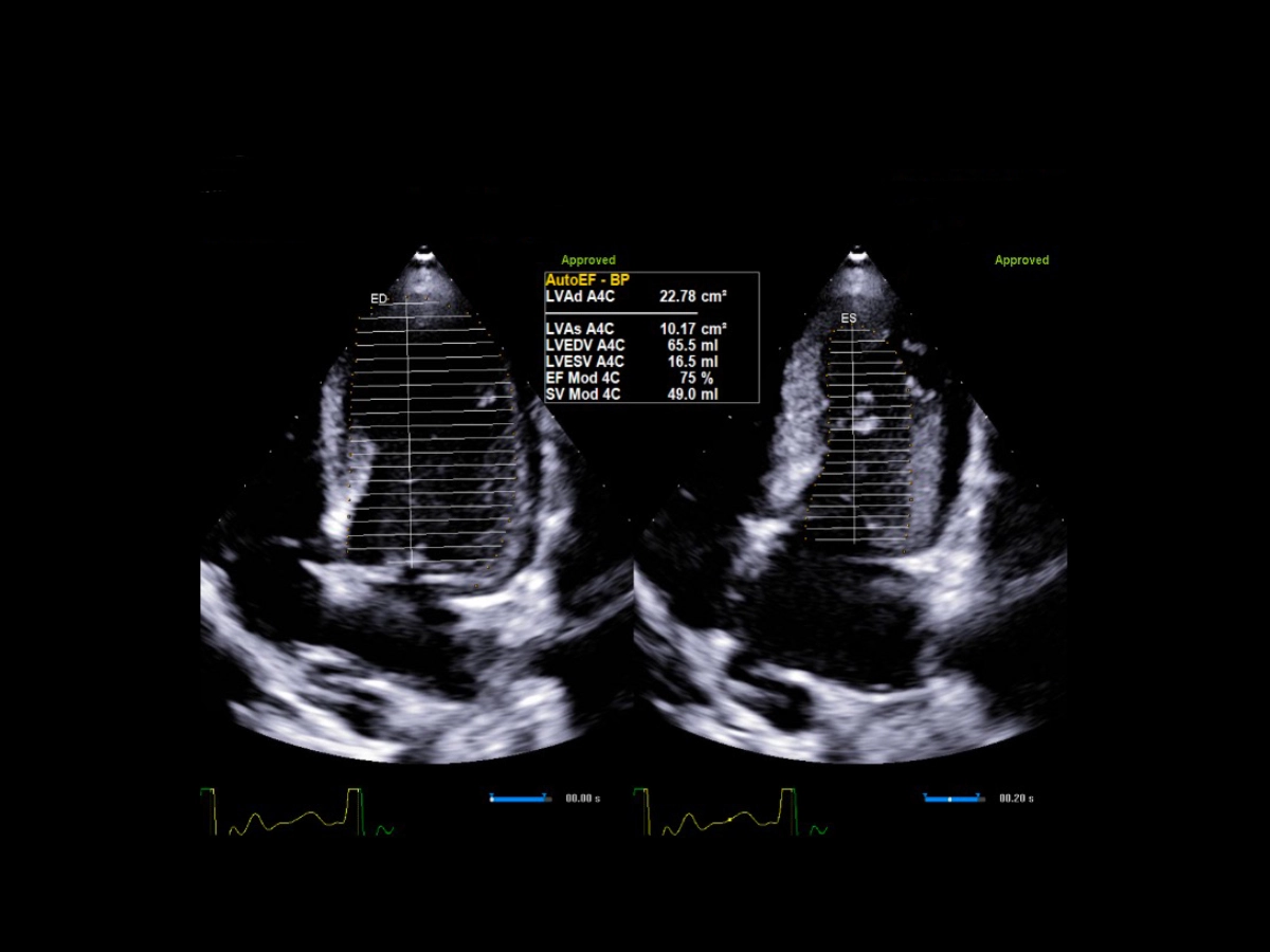
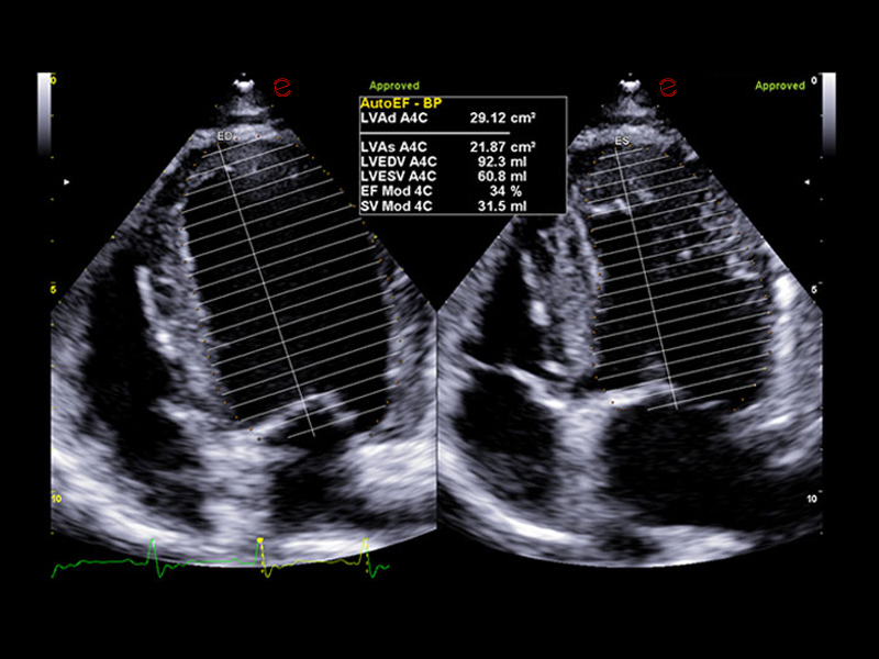
Esaote, leveraging Zero-click Technology supported by Artificial Intelligence (A.I.), offers AutoEF measurement, which automatically calculates values of the left ventricle during both systolic and diastolic phases, thereby calculating the Ejection Fraction (EF). This not only saves time in your workflow but also increases confidence in your assessments. Within seconds, the LV volumes and the corresponding EF are displayed.
Advanced cardiac assessment for a more precise diagnosis
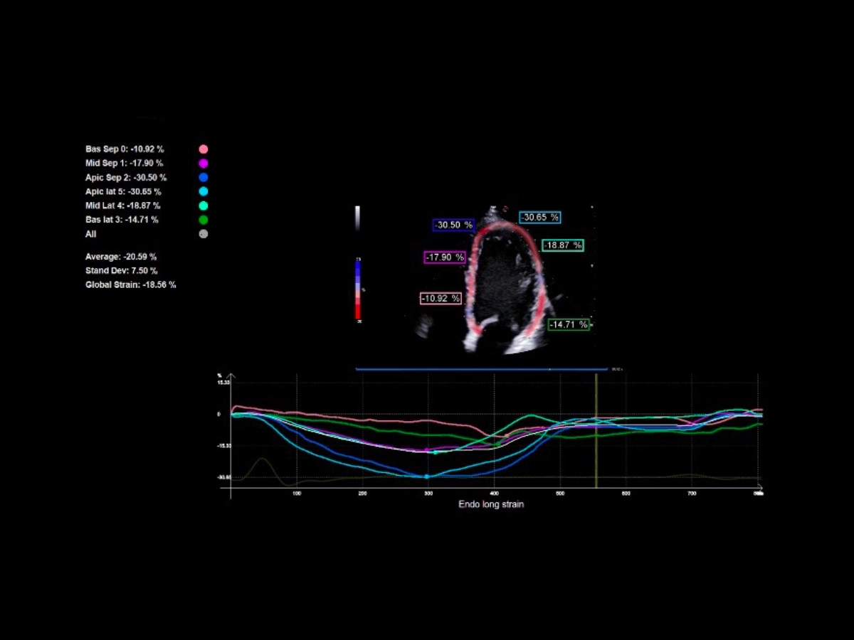
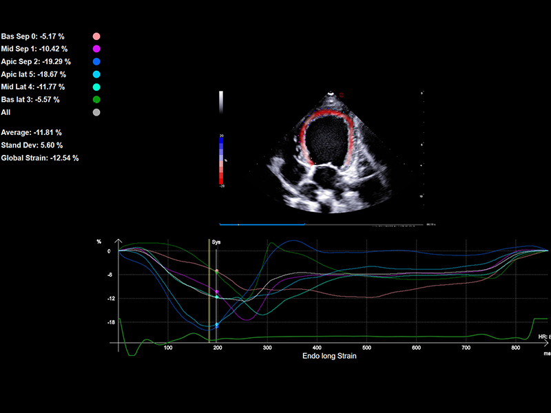
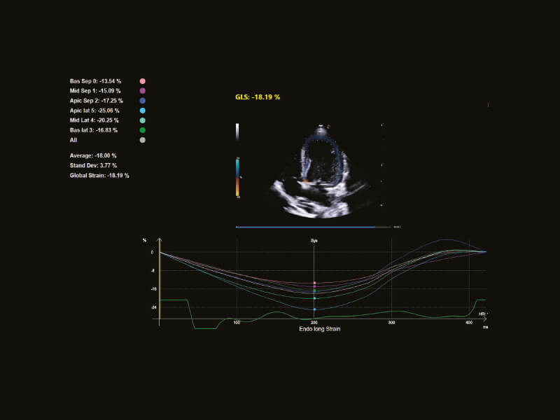
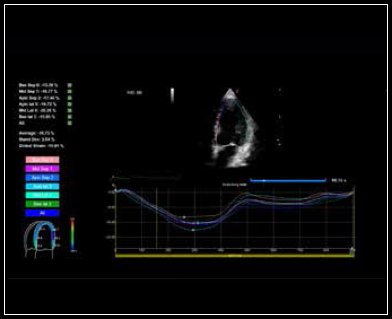
Echocardiographic Strain enables differentiation between passive and active movements of the cardiac muscle. Esaote XStrain™ is an advanced technology powered by A.I. that, with just one click, detects and tracks endocardial borders to provide Global Longitudinal Strain (GLS) and Strain values for each segment. Additionally, Esaote XStrain™ allows for a clear representation of values for each left ventricular segment using a Bull’s Eye layout. Furthermore, aside from LV Strain assessment, Esaote offers the ability to evaluate strain in the right ventricle and right atrium.
Clinical Images
Related systems
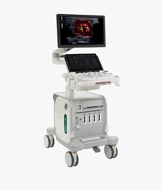
MyLab™X90VET
Premium VET system with new technology of probes and an extended package of advanced tools powered by A.I. to fit with the needs of expert customers.

MyLab™X8VET
Performant VET system which offers comfort and high level of information in a wide range of applications.
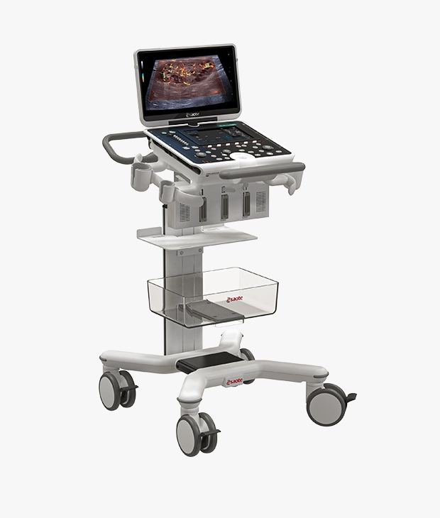
MyLab™Omega eXP VET
Top-class portable VET system which offers an extraordinary user experience in the daily routine.
Technology and features are device/configuration-dependent. Specifications subject to change without notice. Information might refer to products or modalities not yet approved in all countries. Product images are for illustrative purposes only. For further details, please contact your Esaote sales representative.
