Orthopedic MRI in small animals
Magnetic Resonance Imaging (MRI) is increasingly becoming a standard method for imaging joints in companion animals, standing alongside other conventional imaging modalities. For instance, the knee region is the most frequently investigated area with MRI, enabling effective imaging of structures such as cranial and caudal cruciate ligaments, menisci, lateral ligaments, intra articular fluid volume, infrapatellar fat pads, and the chondral surface of articular bones1.

The unique capabilities of MRI, such as its excellent soft tissue contrast and multiplanar imaging, contribute to its growing popularity in musculoskeletal assessments. This non-ionizing and non-invasive technique provides detailed insights into joint structures, aiding in the accurate diagnosis of various conditions affecting companion animals.
Beyond aiding in diagnosis, MRI equips clinicians with valuable insights, allowing for the prediction of prognosis and, in certain instances, aiding in the establishment of treatment plans. The multifaceted utility of MRI in providing detailed anatomical and pathological information contributes significantly to the comprehensive understanding and management of musculoskeletal issues in companion animals.
Esaote's dedicated MRI VET line exemplifies a commitment to advancing veterinary medicine through technologically sophisticated solutions. Tailored specifically for the anatomical idiosyncrasies of veterinary patients, this specialized equipment ensures optimal imaging quality and diagnostic precision.
INTERVIEWS
Testimonials

VIDEO
Interview on MRI veterinary imaging
Dr. Martin Konar

VIDEO
Enhancing clinical practice with Esaote Vet-MR Grande
Dr. Aleksandrs Ozols
Dedicated VET workflow
The attainment of high-quality imaging in veterinary MRI necessitates specific scan protocols and meticulous workflows. Esaote's long standing collaboration with private veterinary clinics and universities, spanning over two decades, has been instrumental in crafting specialized designs and user interfaces tailored for veterinary practice.

Esaote's MRI systems are equipped with an extensive library of protocols, meticulously developed in collaboration with esteemed veterinary professionals. These protocols cater to animals of varying types and sizes, ensuring comprehensive coverage from the smallest to the largest pets. Veterinarians benefit from a specific set of sequences designed to facilitate swift examinations and high throughput, addressing a diverse range of pathologies in the most efficient manner.
In veterinary imaging, Esaote prioritizes a streamlined workflow for enhanced efficiency in daily activities. The comprehensive veterinary design includes a diverse array of receiving coils in various sizes and shapes. These coils are meticulously crafted to boost the signal-to-noise ratio, ensuring heightened resolution without any compromise. This technical refinement enables clinicians to attain precise diagnoses, providing valuable insights into prognosis and facilitating potential surgical planning.

Beyond hardware enhancements, Esaote places equal emphasis on the veterinary user interface. This interface is thoughtfully designed to enhance user familiarity, ultimately minimizing investigation time.

Joints MRI examination
The MRI examination of animal joints is pivotal for diagnosing various pathologies within the musculoskeletal system.
Magnetic resonance methods in animals joint imaging utilize standard sequences including high-resolution T1-weighted, T2-weighted, Short Tau Inversion Recovery (STIR), and fat-suppressed proton density. Additional sequences like Gradient Echo (GE) are applied for visualizing ligament structures and tendons, while 3D sequences such as 3D SST1 and 3D SHARC are particularly beneficial in MRI diagnostics. These sequences offer high tissue contrast and provide accurate and sensitive data for examining bone tissue, cartilage, and articular fluid.
Esaote systems go a step further by incorporating a diverse range of Fat Suppression sequences, including STIR, XBONE, and SPED, enhancing the identification of traumatic lesions and edema effectively.
Overall, MRI's precision in joint imaging supports thorough diagnostic evaluations and informs targeted treatment strategies for musculoskeletal conditions in animals.

References
- Adamiak Z, Jaskólska M, Matyjasik H, Pomianowski A, Kwiatkowska M. Magnetic resonance imaging of selected limb joints in dogs. Pol J Vet Sci. 2011;14(3):501-5. doi: 10.2478/v10181-011-0075-y. PMID: 21957749.
Magnetic Resonance systems for orthopedic in small animals
Magnifico Vet
The latest permanent MRI system from Esaote, the Magnifico Vet combines cutting-edge technology with a permanent magnet to deliver top-tier diagnostic quality at an affordable cost.
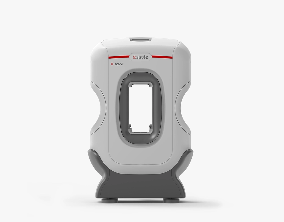
O-scan VET
This customer-driven MRI system is enhanced with vet-exclusive technology developed to better serve veterinary clinics to improve diagnostic efficacy and daily workflows.
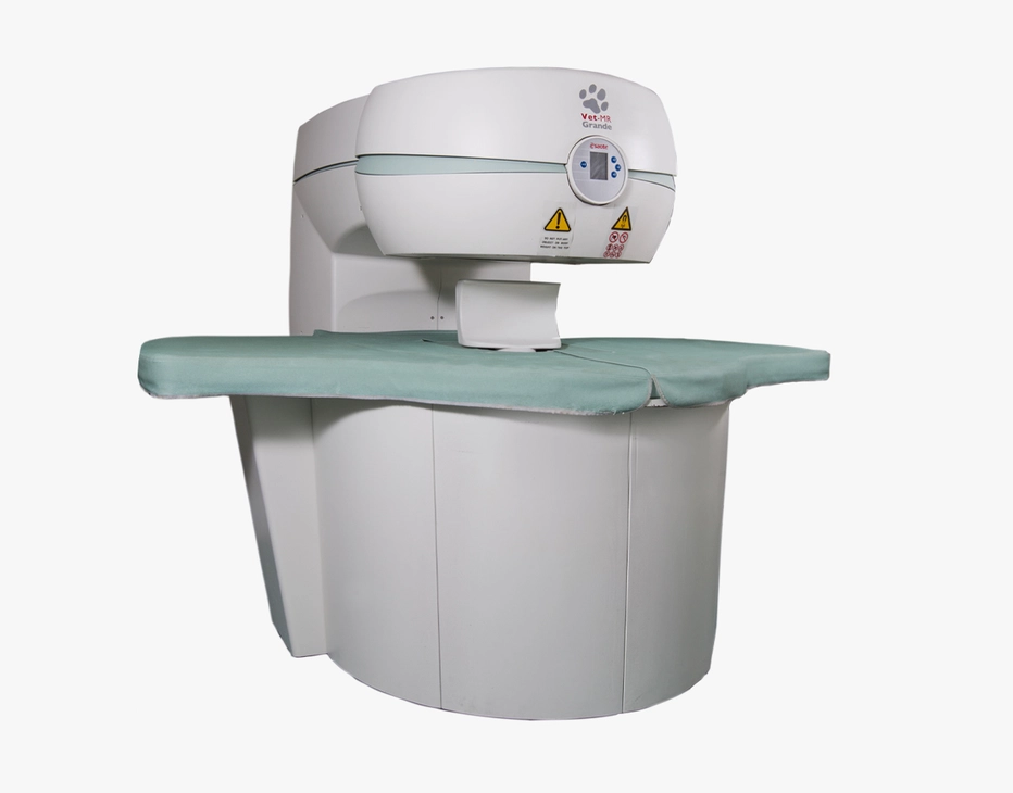
Vet-MR Grande
The innovative MRI scanner is tailored specifically for veterinary use with technology developed to enhance image quality and diagnostic capabilities.
Technology and features are device/configuration-dependent. Specifications subject to change without notice. Information might refer to products or modalities not yet approved in all countries. Product images are for illustrative purposes only. For further details, please contact your Esaote sales representative.


