Neuroimaging MRI in small animals
Magnetic Resonance Imaging (MRI) has established itself as the preeminent imaging modality in veterinary neurology and neurovascular diagnostics. This non-invasive and non-ionizing technique provides unparalleled soft tissue resolution, rendering it indispensable for the precise examination of the nervous system in companion animals.

Scientific investigations have consistently demonstrated the pivotal role of MRI in diagnosing a spectrum of neurological conditions, offering clinicians unparalleled insights into the pathophysiological aspects of these diseases1,2. Moreover, the modality is not only diagnostically potent but also serves as a prognostic and therapeutic tool, furnishing clinicians with imperative data to predict outcomes and formulate tailored treatment strategies.
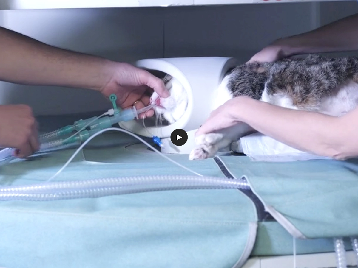
Esaote's dedicated MRI VET line exemplifies a commitment to advancing veterinary medicine through technologically sophisticated solutions. Tailored specifically for the anatomical idiosyncrasies of veterinary patients, this specialized equipment ensures optimal imaging quality and diagnostic precision in the realm of veterinary neuroimaging.
INTERVIEWS
Testimonials

VIDEO
The impact of Vet-MR Grande on Neurology and Surgery
Dr. Beatrix Nanai

VIDEO
Enhancing clinical practice with Esaote Vet-MR Grande
Dr. Aleksandrs Ozols

VIDEO
Interview on MRI veterinary imaging
Dr. Konrad Jurina
Dedicated VET workflow
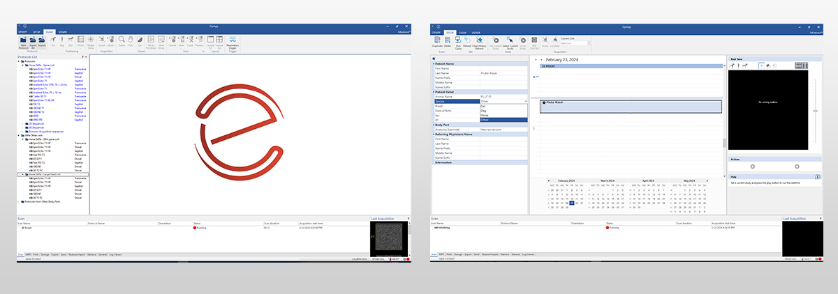
The attainment of high-quality imaging in veterinary MRI necessitates specific scan protocols and meticulous workflows. Esaote's long standing collaboration with private veterinary clinics and universities, spanning over two decades, has been instrumental in crafting specialized designs and user interfaces tailored for veterinary practice.
Esaote's MRI systems are equipped with an extensive library of protocols, meticulously developed in collaboration with esteemed veterinary professionals. These protocols cater to animals of varying types and sizes, ensuring comprehensive coverage from the smallest to the largest pets. Veterinarians benefit from a specific set of sequences designed to facilitate swift examinations and high throughput, addressing a diverse range of pathologies in the most efficient manner.
In veterinary imaging, Esaote prioritizes a streamlined workflow for enhanced efficiency in daily activities. The comprehensive veterinary design includes a diverse array of receiving coils in various sizes and shapes. These coils are meticulously crafted to boost the signal-to-noise ratio, ensuring heightened resolution without any compromise. This technical refinement enables clinicians to attain precise diagnoses, providing valuable insights into prognosis and facilitating potential surgical planning.

Beyond hardware enhancements, Esaote places equal emphasis on the veterinary user interface. This interface is thoughtfully designed to enhance user familiarity, ultimately minimizing investigation time.
MRI Examination of the brain
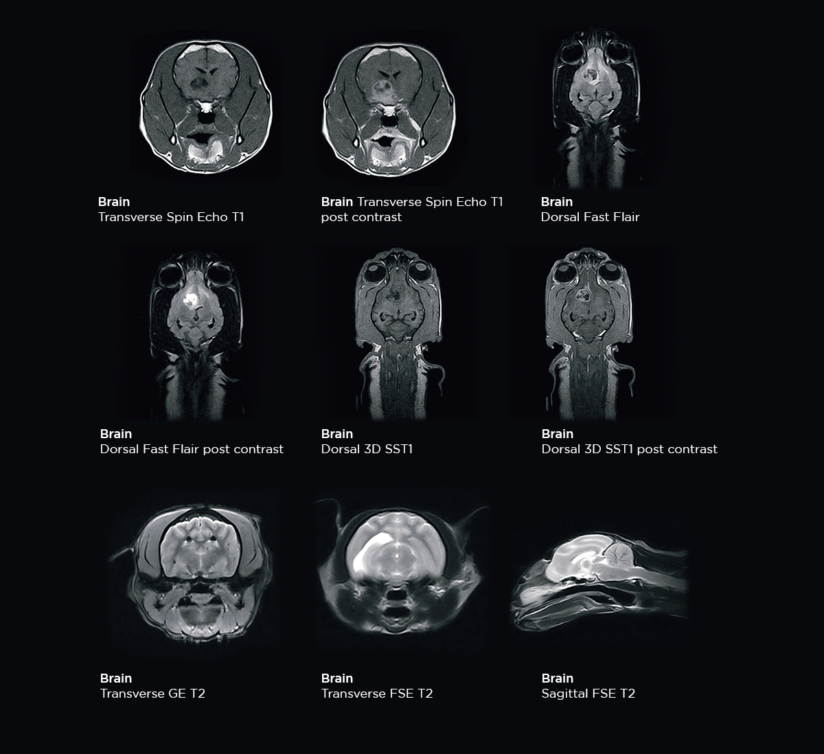
Magnetic Resonance Imaging (MRI) of the neurocranium proves to be a potent tool for evaluating various conditions associated with the brain and neurological system. This encompasses the assessment of both extra and intra-axial intracranial lesions, such as neoplasia, hematoma, cysts, meningioma, and nasal tumors3. A standard basic examination typically includes the execution of T1 and T2-weighted sequences, along with the Fluid Attenuation Inversion Recovery (FLAIR) sequence. The user interface facilitates contrast media management during the imaging process.
Extending head examinations in veterinary MRI involves the utilization of Diffusion-Weighted Imaging (DWI) and Angiographic sequences to explore specific pathologies. These advanced imaging techniques provide additional insights into the microstructural and vascular aspects of the examined tissues, enhancing the diagnostic capabilities for veterinarians.
Esaote veterinary MRI systems demonstrates exceptional proficiency in imaging the head with remarkable anatomical detail, allowing for precise visualization and analysis, ultimately enhancing the clinician's ability to make accurate and informed diagnoses in veterinary medicine.
MRI examination of the spine
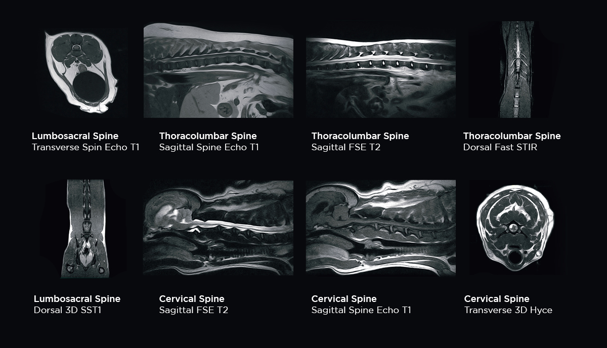
Magnetic Resonance Imaging (MRI) stands as a valuable modality for assessing the spine in animals, offering superior tissue contrast across different planes. This capability aids clinicians in the detection and evaluation of various spine diseases, including disc neoplasia, spondylomyelopathy, and stenosis4. The multi-planar imaging provided by MRI enhances the diagnostic precision, allowing for a comprehensive understanding of spinal conditions in veterinary medicine.
In a routine basic examination of the spine, T1 and T2-weighted sequences, along with Fast Short Time Inversion Recovery (FSTIR), are commonly employed. To augment the examination and improve diagnostic capabilities, the incorporation of 3D volumetric sequences such as 3DSST1 and 3DHYCE is recommended. These sequences can simultaneously scan the anatomy of interest in all three planes with high resolution. Moreover, 3DSST1 is particularly beneficial for both pre- and post-contrast imaging, enhancing the overall diagnostic efficacy of spinal assessments in veterinary medicine.
References
- Joslyn, S. and Hague, D. (2016), Magnetic resonance imaging of the small animal brain. In Practice, 38: 373-385. https://doi.org/10.1136/inp.i4183
- Fuchs J, Domaniža M, Kuricová M, Lipták T, Ledecký V. Comparison of Imaging Methods and Population Pattern in Dogs with Spinal Diseases in Three Periods between 2005 and 2022: A Retrospective Study. Vet Sci. 2023 May 18;10(5):359. doi: 10.3390/vetsci10050359. PMID: 37235442.
- Silke Hecht, William H. Adams, MRI of Brain Disease in Veterinary Patients Part 1: Basic Principles and Congenital Brain Disorders, Veterinary Clinics of North America: Small Animal Practice, Volume 40, Issue 1, 2010, Pages 21-38, https://doi.org/10.1016/j.cvsm.2009.09.005.
- da Costa RC, Samii VF. Advanced imaging of the spine in small animals. Vet Clin North Am Small Anim Pract. 2010 Sep;40(5):765-90. doi: 10.1016/j.cvsm.2010.05.002
Magnetic Resonance systems for neuroimaging in small animals
Magnifico Vet
The latest permanent MRI system from Esaote, the Magnifico Vet combines cutting-edge technology with a permanent magnet to deliver top-tier diagnostic quality at an affordable cost.
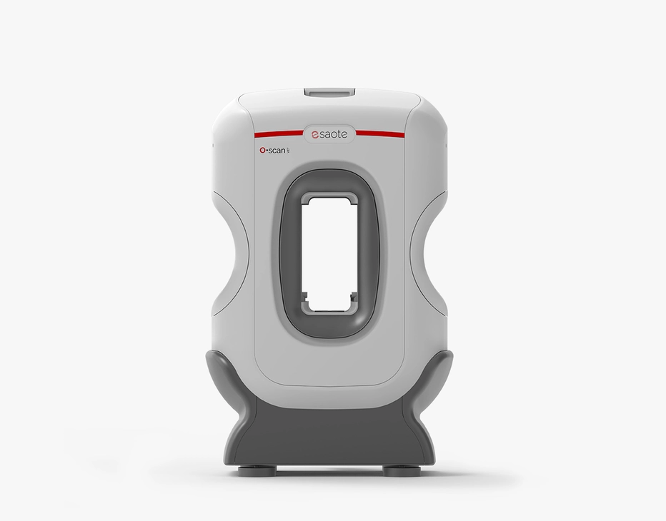
O-scan VET
This customer-driven MRI system is enhanced with vet-exclusive technology developed to better serve veterinary clinics to improve diagnostic efficacy and daily workflows.
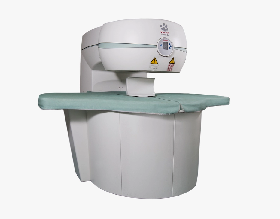
Vet-MR Grande
The innovative MRI scanner is tailored specifically for veterinary use with technology developed to enhance image quality and diagnostic capabilities.
Technology and features are device/configuration-dependent. Specifications subject to change without notice. Information might refer to products or modalities not yet approved in all countries. Product images are for illustrative purposes only. For further details, please contact your Esaote sales representative.

