Orthopedic MRI in equine
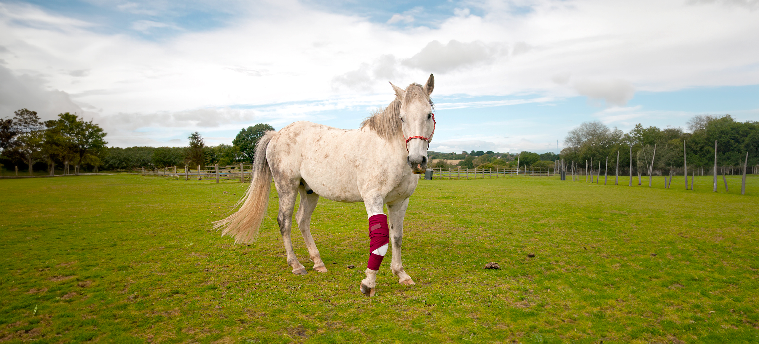
Magnetic Resonance Imaging (MRI) stands as the gold standard for detecting lameness in horses1. It plays a pivotal role in the accurate diagnosis of lesions, distinguishing between joint conditions and tendon/ligament issues. This precision is crucial for formulating effective treatment plans and rehabilitation protocols, ultimately establishing a valid prognosis.
In the realm of bone disorders, MRI serves as the reference technique, unrivaled in identifying and documenting lesions that may escape routine imaging methods. Notably, MRI excels in highlighting bone marrow edema-like (BMEL) lesions, which remain undetectable with CT, even with contrast medium injection. In cases of joint, tendon, and ligament disorders, MRI complements radiography and ultrasonography, providing valuable additional information. While it does not replace these techniques, MRI's high complementarity ensures a more comprehensive and accurate diagnosis.
The safety profile of MRI, being non-ionizing and non-invasive, positions it as a highly favorable medical procedure for sport and racehorses. Esaote's commitment to veterinary medicine is evident through its dedicated MRI VET line, designed to provide optimal solutions for various clinical applications in equine care, further advancing diagnostic capabilities for horses.


Dedicated VET workflow
Achieving good image quality in veterinary MRI relies heavily on specific scan protocols and precise workflows. Esaote has been at the forefront of this endeavor, collaborating with private veterinary clinics and university sites for over two decades. This long standing collaboration has been instrumental in developing a dedicated design and user interface tailored for veterinary practice.
The examination protocol is contingent upon the anatomical region under investigation and the nature of suspected or identified lesions, distinguishing between joint conditions and tendon/ligament issues. To streamline and optimize the imaging process, Esaote MRI systems are equipped with a comprehensive library of protocols. These protocols, developed in collaboration with esteemed veterinary experts, ensure not only fast examinations but also high throughput across various pathologies. This commitment to tailored protocols enhances the efficiency and diagnostic accuracy of veterinary MRI, ultimately benefiting both clinicians and their animal patients.
In veterinary imaging, Esaote prioritizes a streamlined workflow for enhanced efficiency in daily activities. The comprehensive veterinary design includes a diverse array of receiving coils in various sizes and shapes. These coils are meticulously crafted to boost the signal-to-noise ratio, ensuring heightened resolution without any compromise. This technical refinement enables clinicians to attain precise diagnoses, providing valuable insights into prognosis and facilitating potential surgical planning.
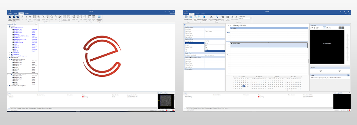
Beyond hardware enhancements, Esaote places equal emphasis on the veterinary user interface. This interface is thoughtfully designed to enhance user familiarity, ultimately minimizing investigation time.
Joints MRI examination
Magnetic Resonance Imaging (MRI) has solidified its status as the gold standard for examining the musculoskeletal anatomy of horses. This advanced imaging technique enables the rapid and precise localization of damage in both bone and soft tissues. Its ability to differentiate between various tissue types empowers veterinary clinicians to assess and manage a spectrum of orthopedic issues, including lameness, joint diseases, soft tissue injuries, and bone abnormalities.
In the context of equine MRI protocols, standard practices often include sequences such as Gradient Echo T2* and 3DSHARC for optimal cartilage visualization, 3DSST1 for volumetric anatomy imaging, and Fast Spin Echo for Proton Density and T2 imaging of ligaments and muscular structures. Esaote systems further enhance diagnostic capabilities by incorporating a diverse range of Fat Suppression sequences like STIR, XBONE, and SPED, facilitating effective identification of traumatic lesions and edema.
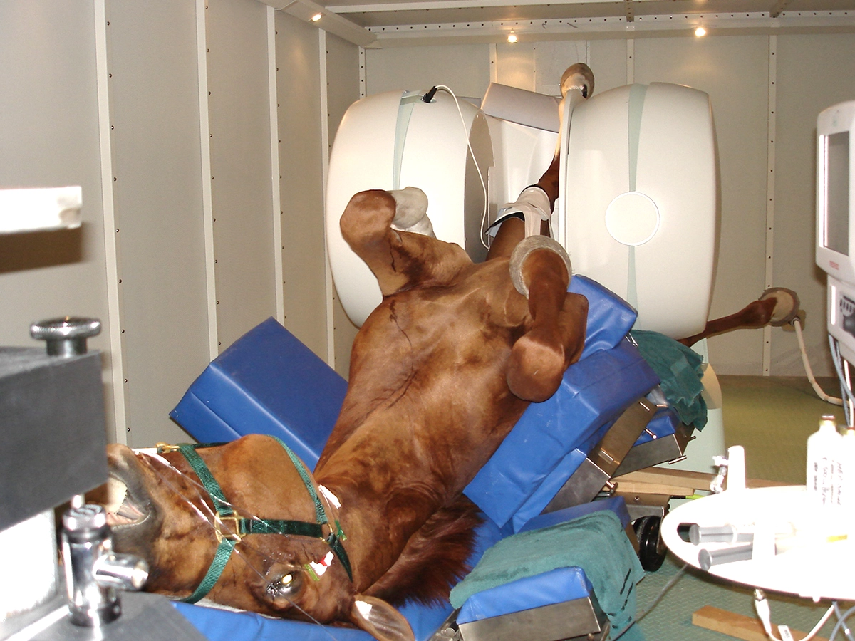
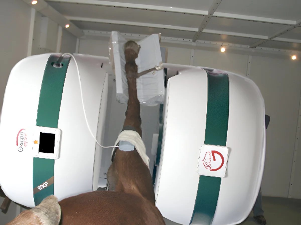
A noteworthy addition to Esaote's offerings is the G-scan Equine, which stands out as the only MRI system providing accurate imaging of the equine stifle by turning the magnet of 90°. Studies have confirmed its efficacy in detecting lesions that initial radiographic and ultrasonographic images may have overlooked2. The G-scan Equine's protocol allows for routine stifle MRI, independent of breed, age, and gender, demonstrating safety and the ability to delineate both bone and soft-tissue pathology in horses.
References
- Barrett MF, Goorchenko GE, Frisbie DD. Comparison of Ultrasound and Magnetic Resonance Imaging for Identifying Soft Tissue Abnormalities in the Palmar Aspect of the Equine Digit. Animals (Basel). 2023 Jul 17;13(14):2328. doi: 10.3390/ani13142328. PMID: 37508105; PMCID: PMC10376038.
- Waselau, M., McKnight, A., & Kasparek, A. (2020). Magnetic resonance imaging of equine stifles: Technique and observations in 76 clinical cases. Equine Veterinary Education. doi:10.1111/eve.13248 10.1111/eve.13248
Magnetic Resonance systems for orthopedic in equine
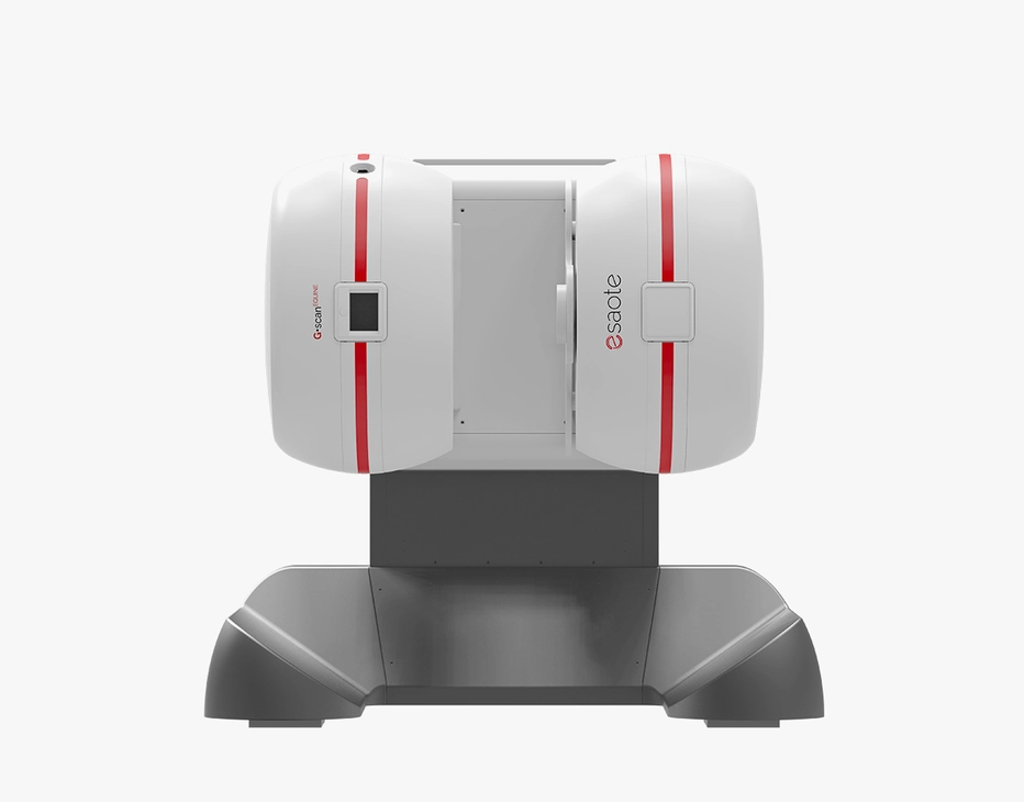
G-scan Equine
The G-scan Equine MRI system is engineered specifically for equine imaging excellence. Its unique design enables seamless patient positioning and accurate investigation of stifle joint issues in horses.
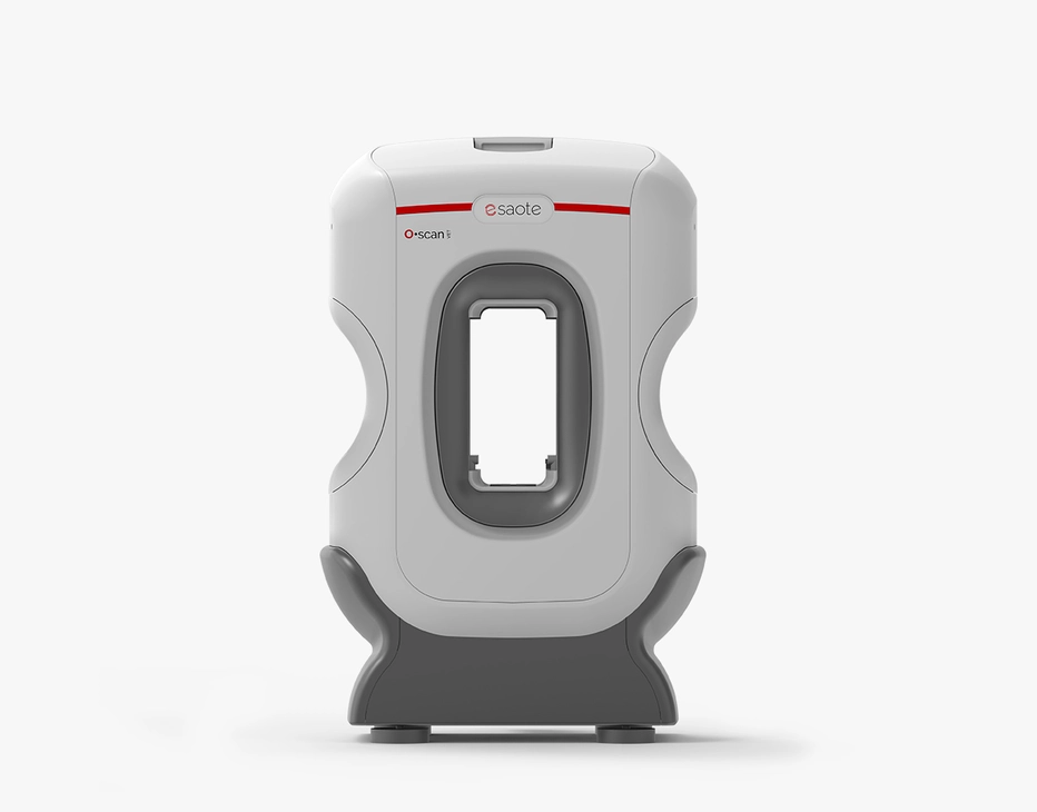
O-scan VET
This customer-driven MRI system is enhanced with vet-exclusive technology developed to better serve veterinary clinics to improve diagnostic efficacy and daily workflows.
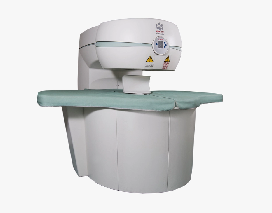
Vet-MR Grande
The innovative MRI scanner is tailored specifically for veterinary use with technology developed to enhance image quality and diagnostic capabilities.
Technology and features are device/configuration-dependent. Specifications subject to change without notice. Information might refer to products or modalities not yet approved in all countries. Product images are for illustrative purposes only. For further details, please contact your Esaote sales representative.
