Neuroimaging MRI in equine
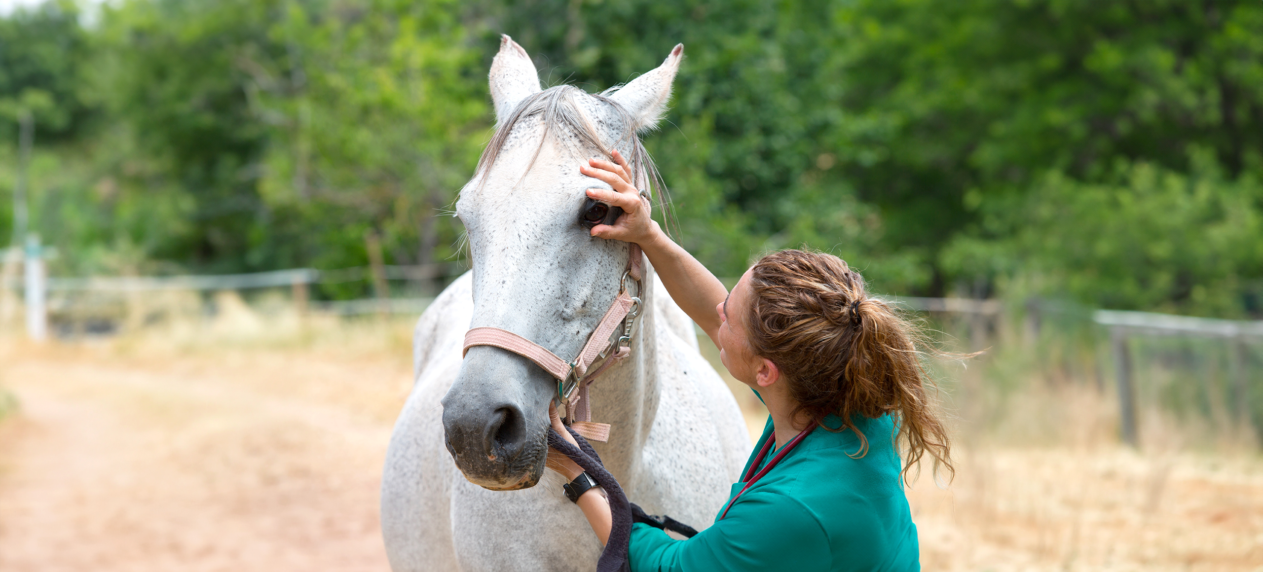
The equine head is a highly complex anatomical region, which is why advanced tomographic imaging techniques, such as computed tomography or magnetic resonance imaging (MRI), are frequently needed for accurate diagnosis and treatment planning.
Neuroimaging MRI, specifically focused on the brain and neurologic system, proves invaluable in providing clinical insights into various conditions such as tumors, strokes, aneurysms, neurological disorders, and trauma. The versatility of MRI in imaging healthy and pathological tissues with different contrasts positions it as a crucial instrument for assessing neurologic pathologies in horses.
MRI scans, known for their safety due to the non-ionizing and non-invasive nature of the method, are considered a cornerstone in veterinary diagnostics, offering a holistic approach for the well-being of our pets. Esaote's commitment to veterinary medicine is evident through its dedicated MRI VET line, designed to provide optimal solutions for various clinical applications, further advancing the diagnostic capabilities for equine healthcare.
Dedicated VET workflow
The attainment of high-quality imaging in veterinary MRI necessitates specific scan protocols and meticulous workflows. Esaote's long standing collaboration with private veterinary clinics and universities, spanning over two decades, has been instrumental in crafting specialized designs and user interfaces tailored for veterinary practice.
Esaote's MRI systems are equipped with an extensive library of protocols, meticulously developed in collaboration with esteemed veterinary professionals. These protocols cater to animals of varying types and sizes, ensuring comprehensive coverage from the smallest to the largest pets. Veterinarians benefit from a specific set of sequences designed to facilitate swift examinations and high throughput, addressing a diverse range of pathologies in the most efficient manner.
In veterinary imaging, Esaote prioritizes a streamlined workflow for enhanced efficiency in daily activities. The comprehensive veterinary design includes a diverse array of receiving coils in various sizes and shapes. These coils are meticulously crafted to boost the signal-to-noise ratio, ensuring heightened resolution without any compromise. This technical refinement enables clinicians to attain precise diagnoses, providing valuable insights into prognosis and facilitating potential surgical planning.
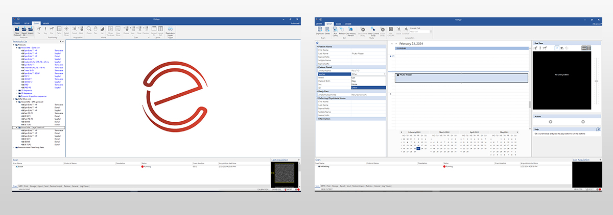
Beyond hardware enhancements, Esaote places equal emphasis on the veterinary user interface. This interface is thoughtfully designed to enhance user familiarity, ultimately minimizing investigation time.
MRI Examination of the brain
Indeed, for neurological cases in both humans and companion animals, MRI has emerged as the preferred diagnostic test due to its superior soft tissue contrast. This non-invasive and non-ionizing technique provides unparalleled resolution, enabling precise examination of the nervous system. The ability of MRI to distinguish grey and white matter regions, along with components of the ventricular system, makes it an indispensable tool for neurological assessments in companion animals.
When compared to the use of MRI to detect appendicular skeletal lesions in horses, equine skull MRI represents an area of limited growth. While using MRI to examine the equine head can be a challenging process, it consistently provides beneficial diagnostic information to the patient and treating clinician.
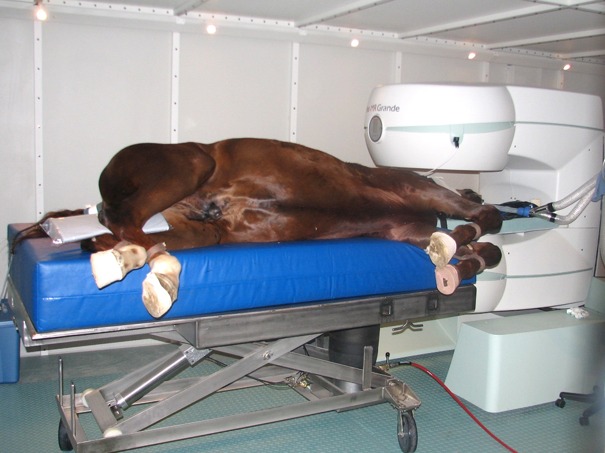
Esaote veterinary systems, such as G-scan Equine and Vet-MR Grande, offer a comprehensive range of sequences to address clinical questions and support veterinarians in diagnosing neurological disorders in horses. A standard neuro equine examination typically involves T1 and T2 weighted sequences, along with FLAIR (Fluid Attenuation Inversion Recovery), with contrast media management facilitated by the user interface. These systems contribute to proficient imaging of the equine head with high anatomical detail, providing a comfortable setting that supports clinicians in the diagnostic process.

Magnetic Resonance systems for neuroimaging in equine
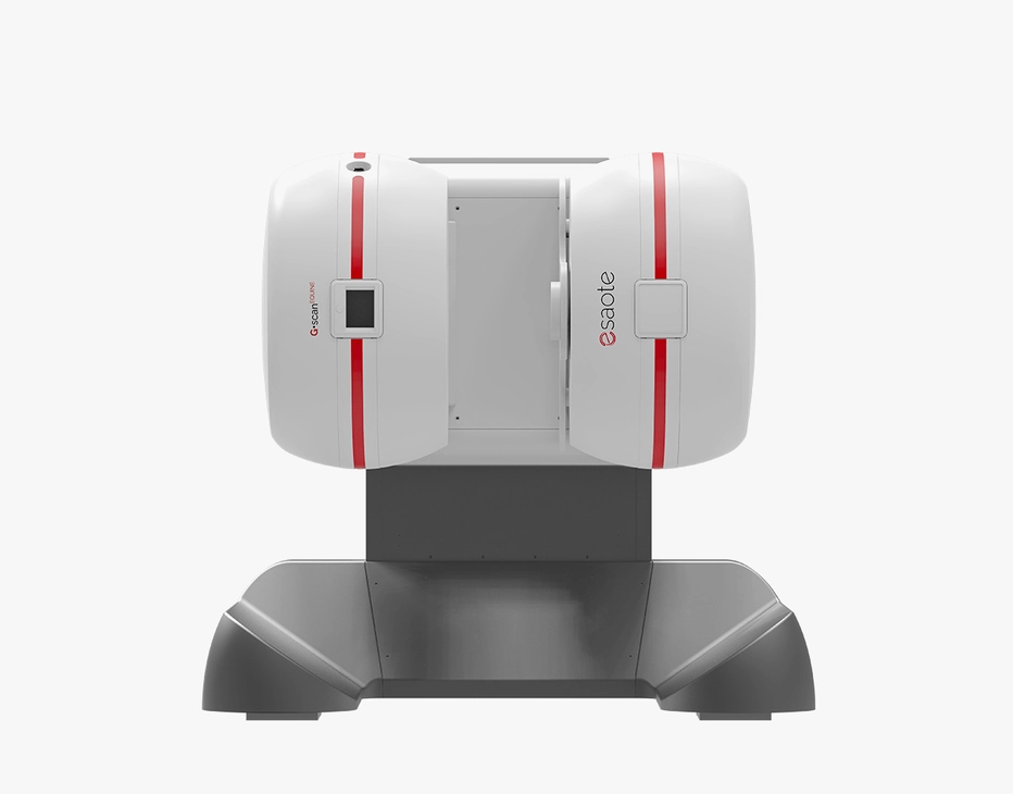
G-scan Equine
The G-scan Equine MRI system is engineered specifically for equine imaging excellence. Its unique design enables seamless patient positioning and accurate investigation of stifle joint issues in horses.
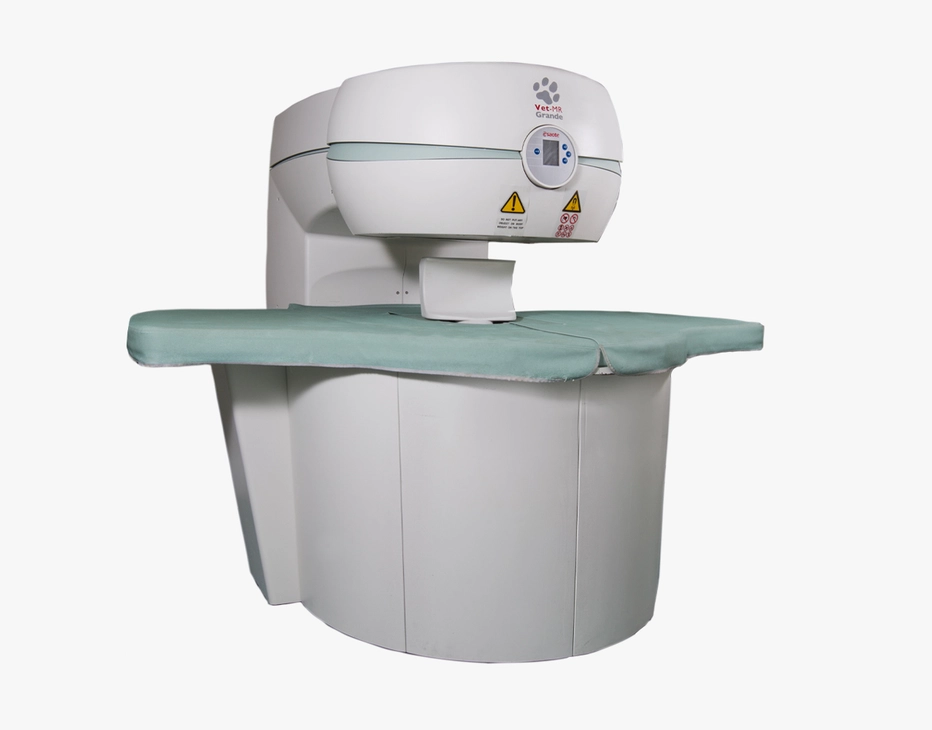
Vet-MR Grande
The innovative MRI scanner is tailored specifically for veterinary use with technology developed to enhance image quality and diagnostic capabilities.
Technology and features are device/configuration-dependent. Specifications subject to change without notice. Information might refer to products or modalities not yet approved in all countries. Product images are for illustrative purposes only. For further details, please contact your Esaote sales representative.
