Tetralogy of fallot
Identification and history
- Name: Goldrake
- Report and medical history: Dog, Corso, male, 3 months.
Echocardiographic evaluation requested by referring veterinarian for suspected congenital heart disease. The patient showed bilateral precordial thrill and a very intense heart murmur, especially on the right hemithorax at the level of the cardiac apex and on the left hemithorax at the level of the heart base.
Diagnostics
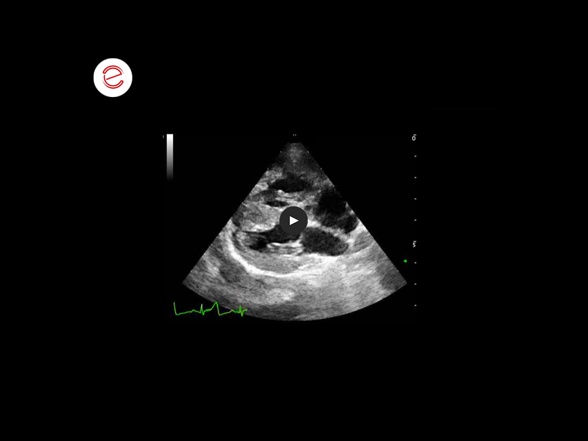
Right parasternal long-axis view of the heart: right ventricle hypertrophy and right atrium dilatation.
Right parasternal long-axis view of the heart: defect of the membranous part of the interventricular septum.
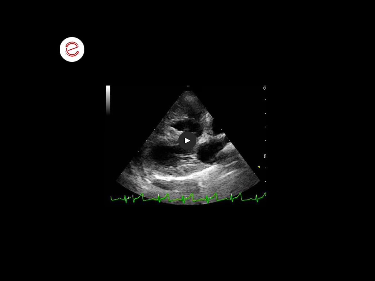
Right parasternal long-axis view of the heart: defect of the membranous part of the interventricular septum and dextroposition of the aorta (aorta overriding the interventricular septum).
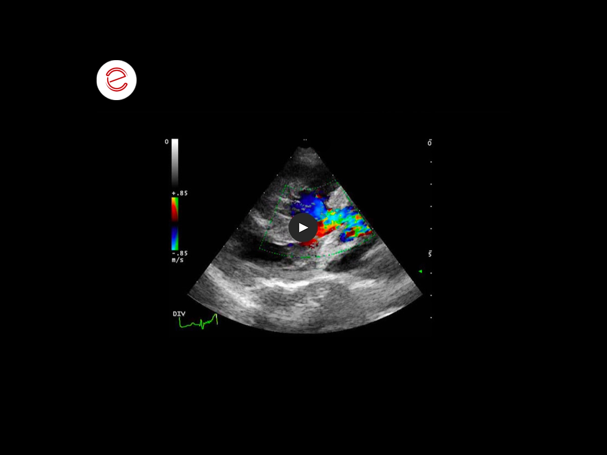
Right parasternal long-axis view of the heart: color Doppler examination highlights turbulent ejection flow within the aorta from both left ventricle and right ventricle (right-to-left shunt).
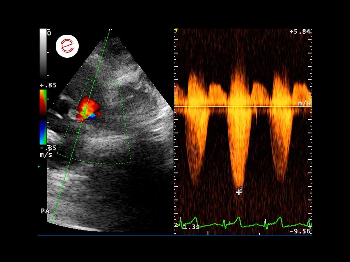
Right parasternal view, continuous wave spectral Doppler: turbulent flow within the pulmonary artery, with a dynamic obstruction component of the right ventricular outflow tract.
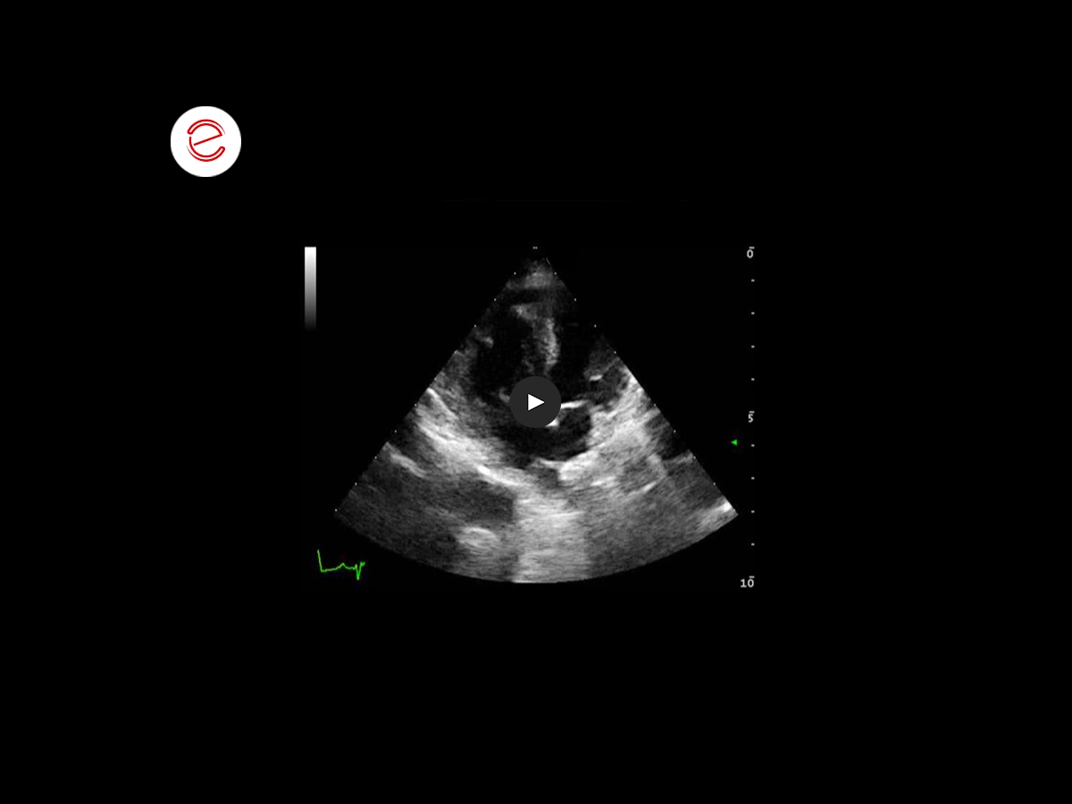
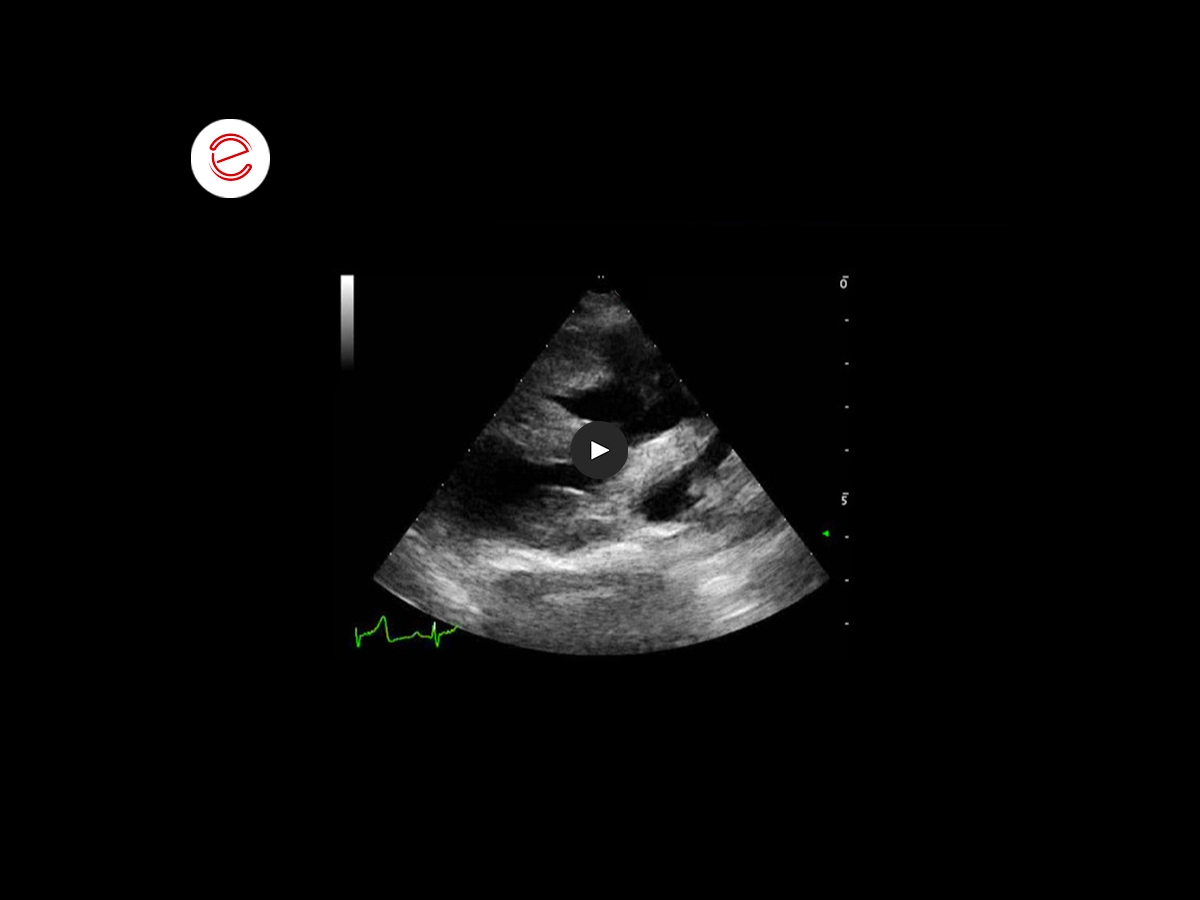
Left apical four-chamber view of the heart: defect of the membranous part of the interventricular septum and defect of the interatrial septum.
Images were acquired using the MyLab™ClassCVET system.
Conclusions and treatment
Echocardiographic Diagnosis: Tetralogy of Fallot, associated with atrial septal defect. The owners did not consider performing surgery to correct the congenital heart disease to be the preferred option, therefore pharmacological treatment and periodic bloodletting therapy were recommended to ensure the patient's hemodynamic balance is as stable as possible.
Alessandro Fruganti, DVM, PhD
University Veterinary Teaching Hospital – University of Camerino (MC), Italy

MyLab is a trademark of Esaote spa.
Technology and features are device/configuration-dependent. Specifications subject to change without notice. Information might refer to products or modalities not yet approved in all countries. Product images are for illustrative purposes only. For further details, please contact your Esaote sales representative.
Other canine clinical cases you may be interested in
Discover the challenges faced, the examinations performed, the solutions adopted, and the treatments recommended.
JUNE 2021
Ventricular septum defect
Claudio Bussadori, DVM, MD, PhD, Dipl. ECVIM (Cardiology)
Clinica Veterinaria Gran Sasso, Milan, Italy
FEBRUARY 2022
Adrenal adenoma
Sergio Fanfoni, DVM, Clinica Veterinaria Santa Cristina
Monte San Savino, Italy, SCIVAC Coordinator
JUNE 2022
Brachial biceps rupture
Laura Martinelli, DVM, MSc
Faculty of Veterinary Medicine, University of Milan, Italy
