Right atrial Hemangiosarcoma
Identification and history
- Name: Toby
- Report and medical history: canine, Yorkshire Terrier, neutered male, 15 years old, 8 kg.
On the day of the visit the patient is developing respiratory distress and in the previous 48 hours, he has presented apathy, anorexia and cough.
Thoracic radiographs show right heart enlargement and unstructured interstitial pulmonary pattern with multiple nodules.
Echocardiography and abdominal ultrasound are requested.
Diagnostics
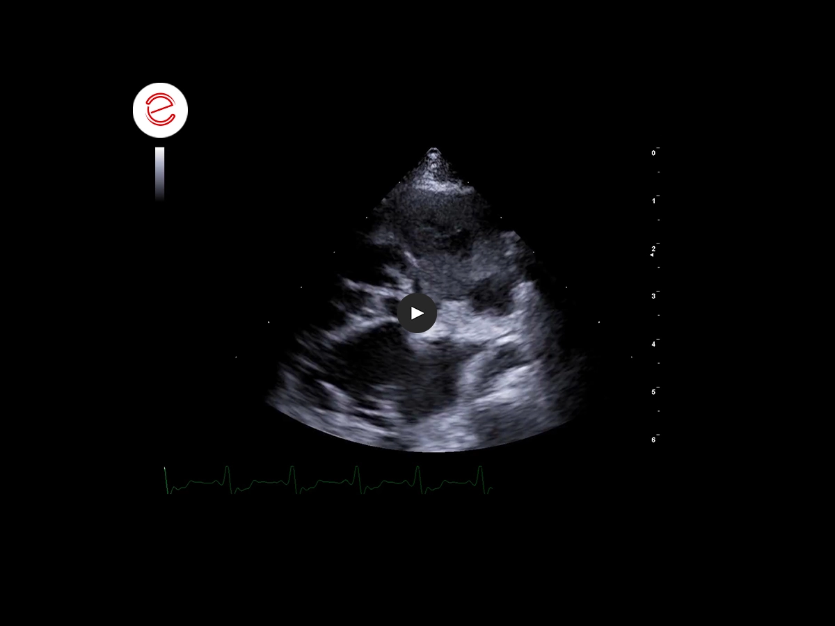
On right parasternal long axis view, optimized for left and right atrium, the presence of a heterogeneous mass adjacent to the right atrium is evidenced. Other findings:
- small right atrium
- thicker mitral valve leaflets
- mild mitral valve regurgitation on color flow
- mild diffuse hypertrophy from the left ventricle
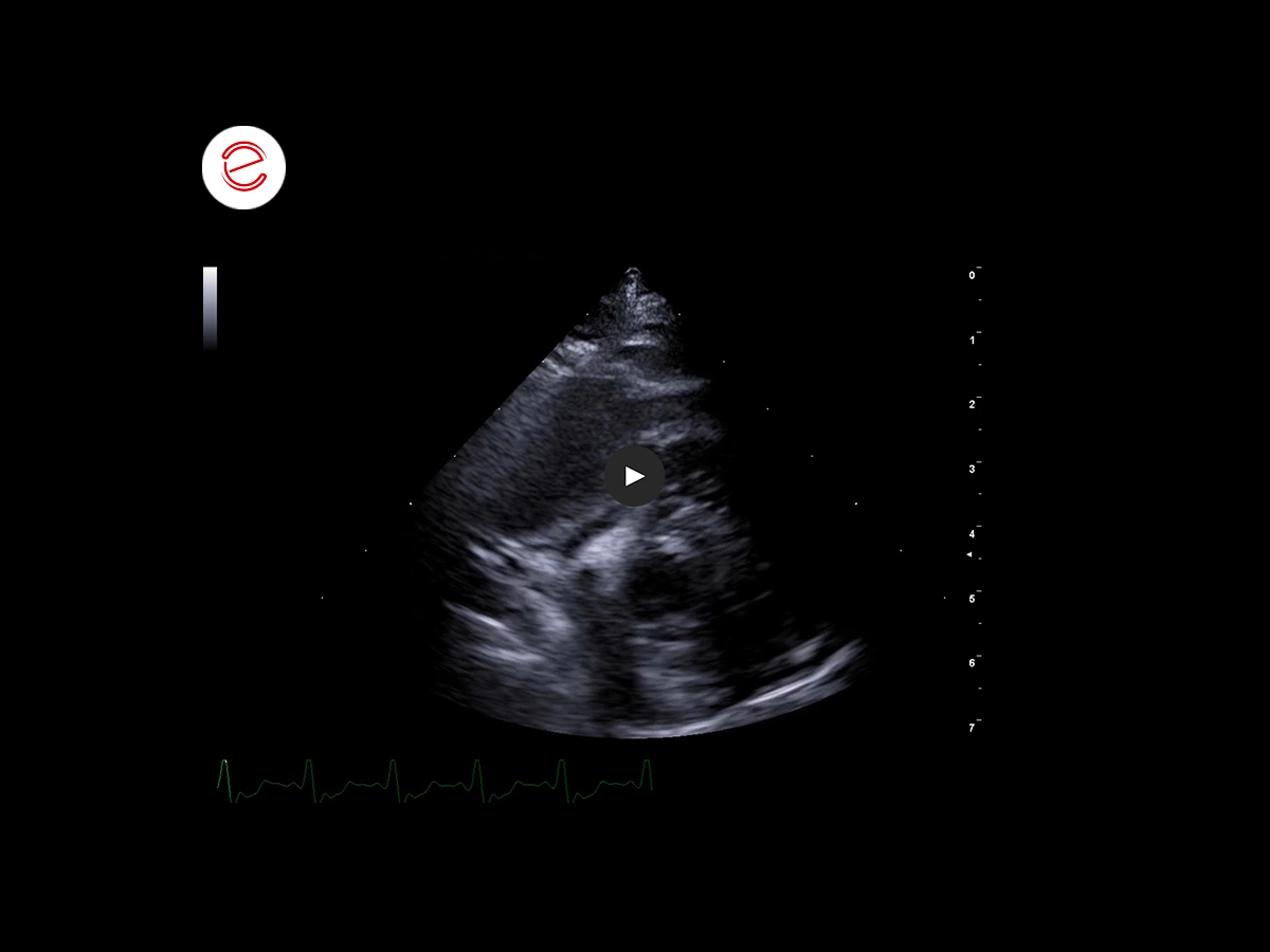
On right parasternal short axis view, at the level of aorta and left atrium, the mass is affecting right atrium free wall and pressing on the cavity.
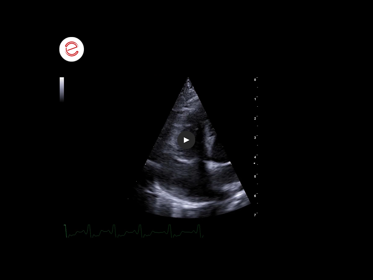
On left apical views, optimized for right heart, the mass is clearly located on the right atrium.
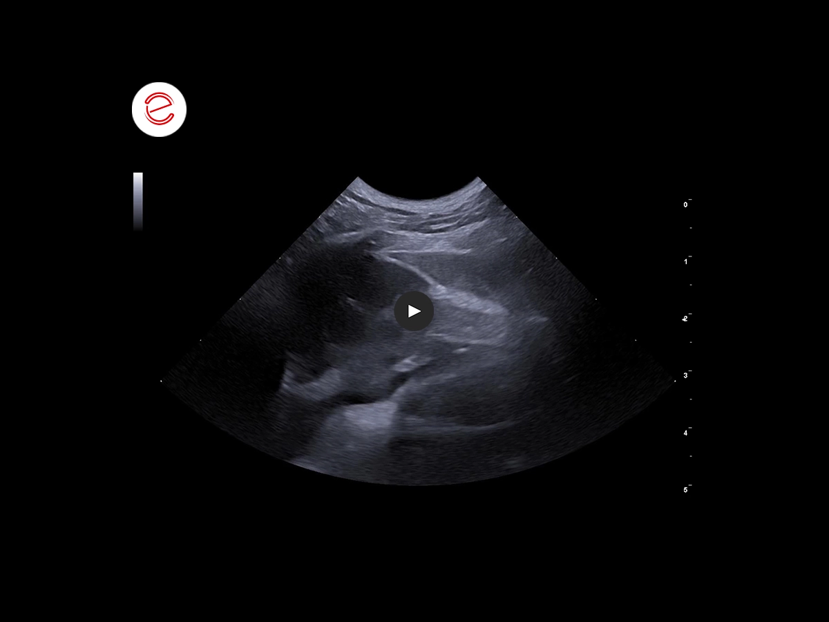
With the thoracic ultrasound the right atrial mass is showing mixed echogenicity with cavitary component.
It is possible to appreciate a thicker pleural line, presence of multiple anechoic nodules and B-lines on the pulmonary parenchyma.

Liver is enlarged, with rounded lobe margins, and multiple heterogeneous nodules are affecting the hepatic parenchyma.
Some pulmonary B-lines are visible across the diaphragm.
Images were acquired using the MyLab™Omega VET system.
Conclusions and treatment
Based on localization and appearance of the cardiac mass, findings are compatible with right atrial hemangiosarcoma and pulmonary and hepatic metastasis. Fine needle aspiration from liver could confirm the diagnosis on this dog. Surgical resection of the mass and chemotherapy can improve survival time on these patients, but prognosis is poor on advanced disease.
Angeles Carrión DVM, Accre. AVEPA in Cardiology. Vetocardia, Veterinary cardiology and ultrasound, Murcia, Spain.

MyLab is a trademark of Esaote spa.
Technology and features are device/configuration-dependent. Specifications subject to change without notice. Information might refer to products or modalities not yet approved in all countries. Product images are for illustrative purposes only. For further details, please contact your Esaote sales representative.
Other canine clinical cases you may be interested in
Discover the challenges faced, the examinations performed, the solutions adopted, and the treatments recommended.

MAY 2025
Adrenomegaly and bilateral nephropathy
University Veterinary Hospital, Department of Diagnostic Imaging, University of Milan, Lodi.

MARCH 2025
Adrenal tumors
Carmelo Marco Bruno, DVM, specialist in pathology and clinic of companion animals, accredited with FSA and BOL

MAY 2024
Synovial Sarcoma
Daniel Sáez, DVM
Centro de Diagnóstico Veterinario Vetpoint®, Chile
