Restrictive cardiomyopathy
Identification and history
- Name: Barbarella
- Report and medical history: Cat, Birman, neutered female, 5 years old.
Echocardiogram request for previous diagnosis of pleural effusion.
Diagnostics
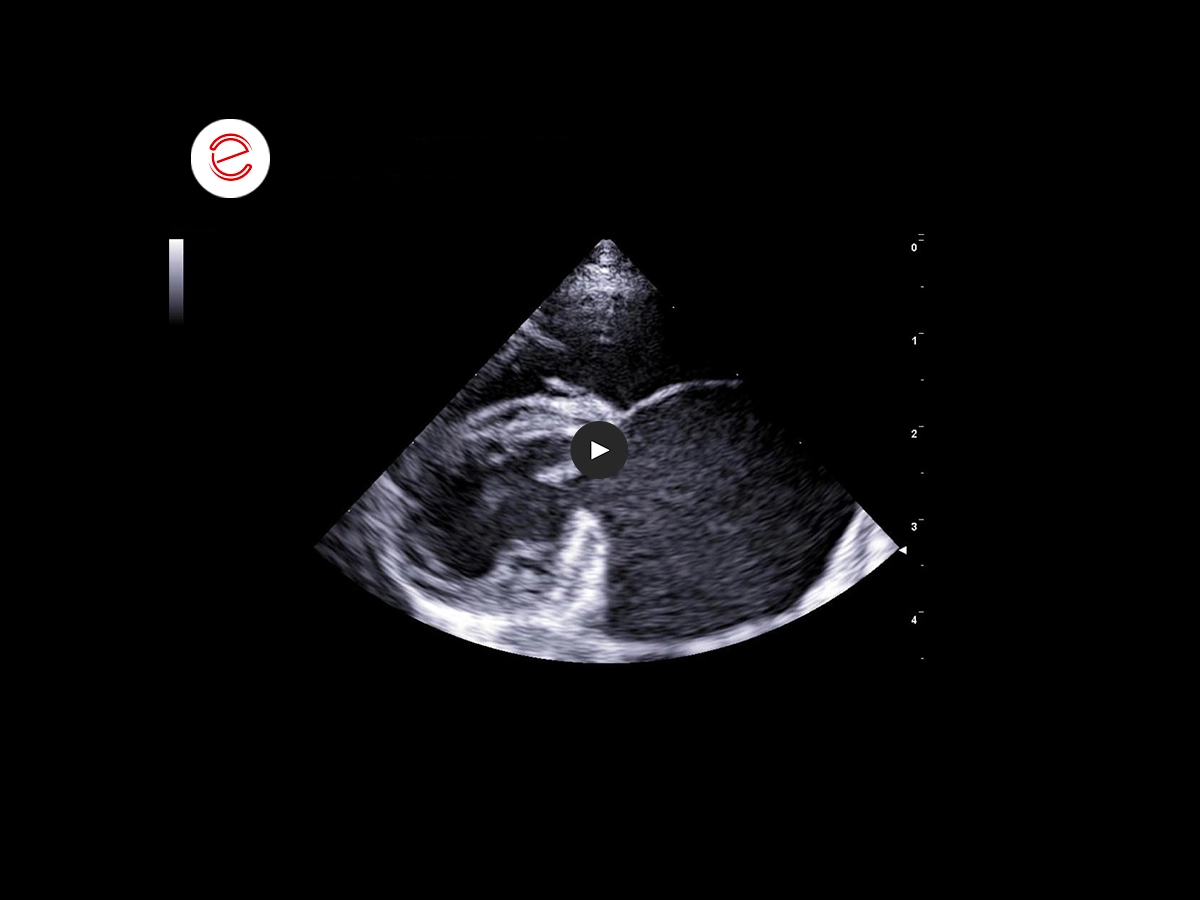
Right parasternal, Standard 1
The echo shows a severe bilateral dilation and signs of “smoke effect” inside the left atrial chamber.
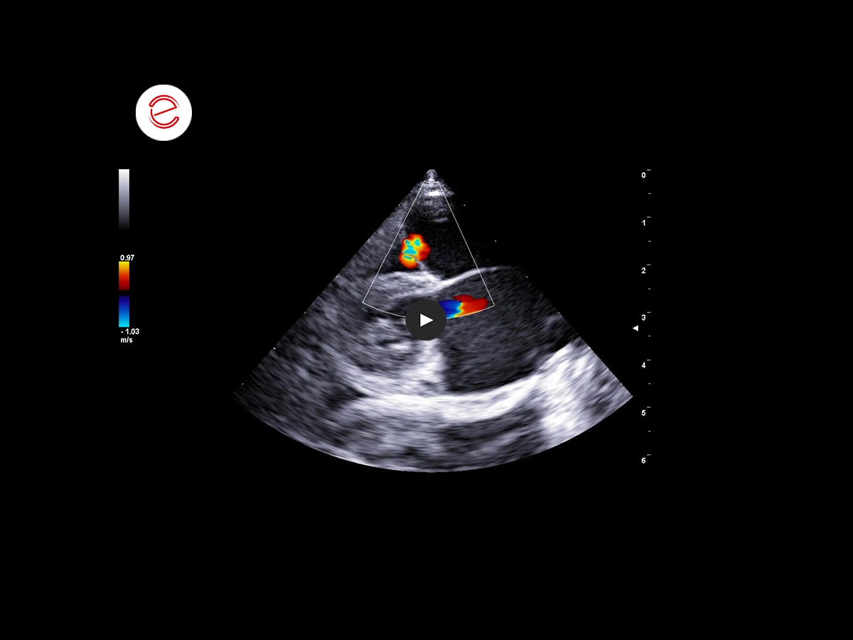
Right parasternal, Standard 1
Colour Doppler shows the presence of a small tricuspid regurgitation jet with an anterior direction.
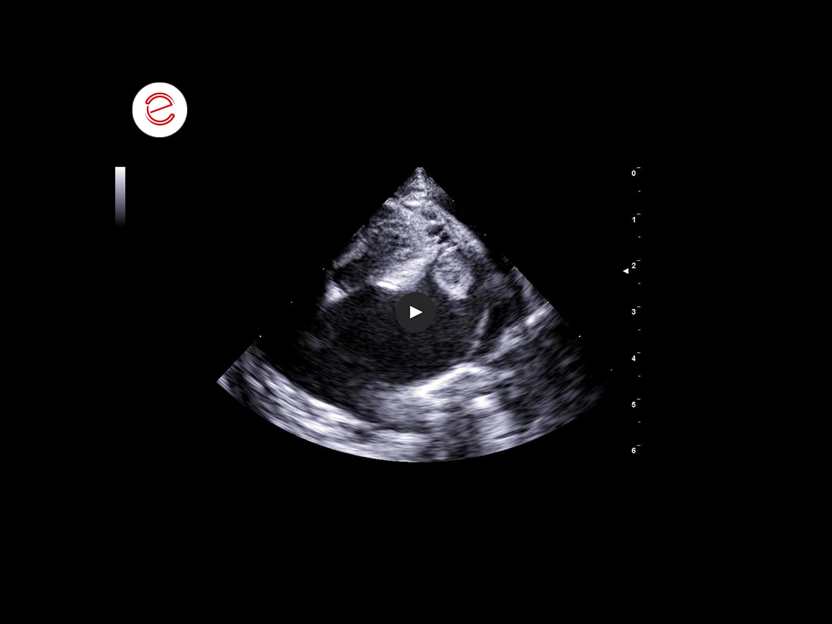
Left parasternal, Apical 4 chamber, optimised for examining the left auricle.
As well as a major increase in the dimensions of the left atrium, there is a marked dilation of the left auricle, inside which a hyperechogenic structure can be seen.
This structure appears to be anchored to the walls of the auricle, but floating in the chamber.

This still image allows measurement of the structure in question and shows its clearly defined margins and rounded, peduncular shape.
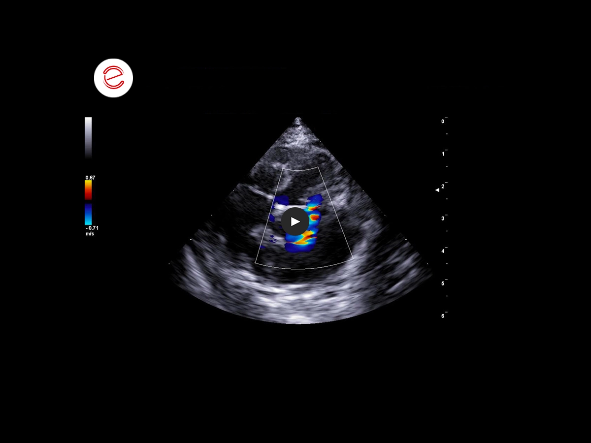
Left parasternal, Apical 4 chamber
Colour Doppler shows a mitral regurgitation jet with a central-anterior direction.
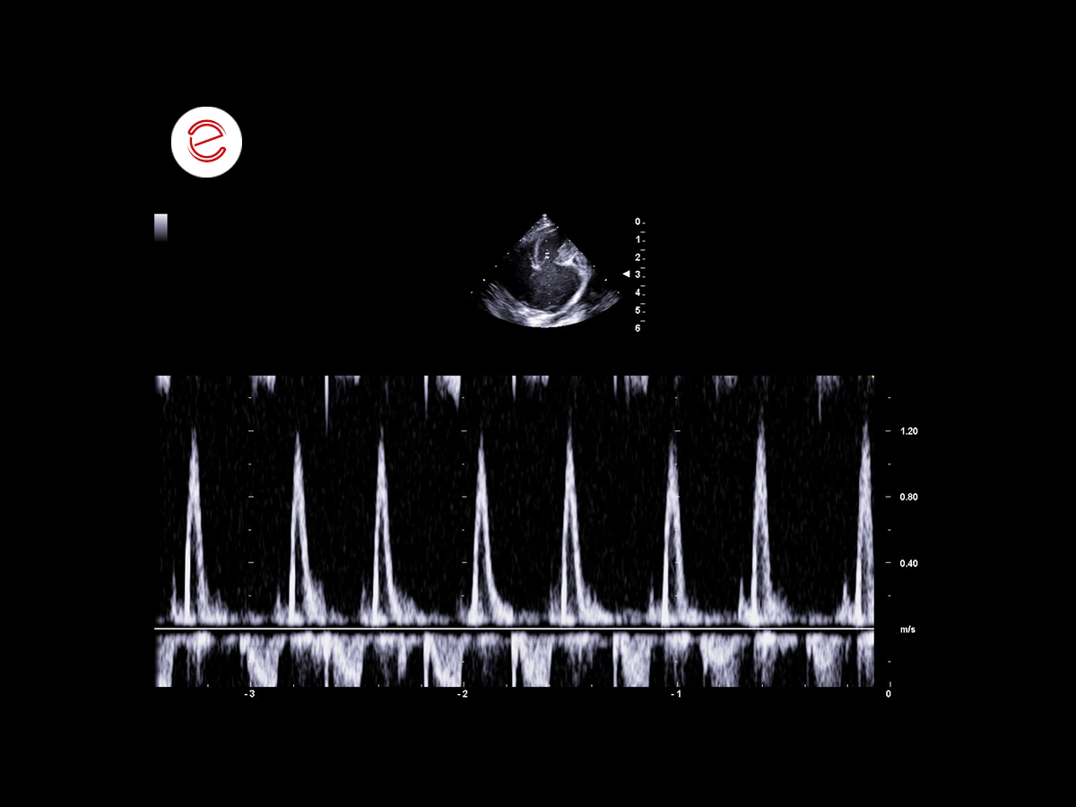
Left parasternal, Apical 4 chamber
Trans-mitral pattern showing just the E-wave, a sign of early ventricular filling at high velocity.
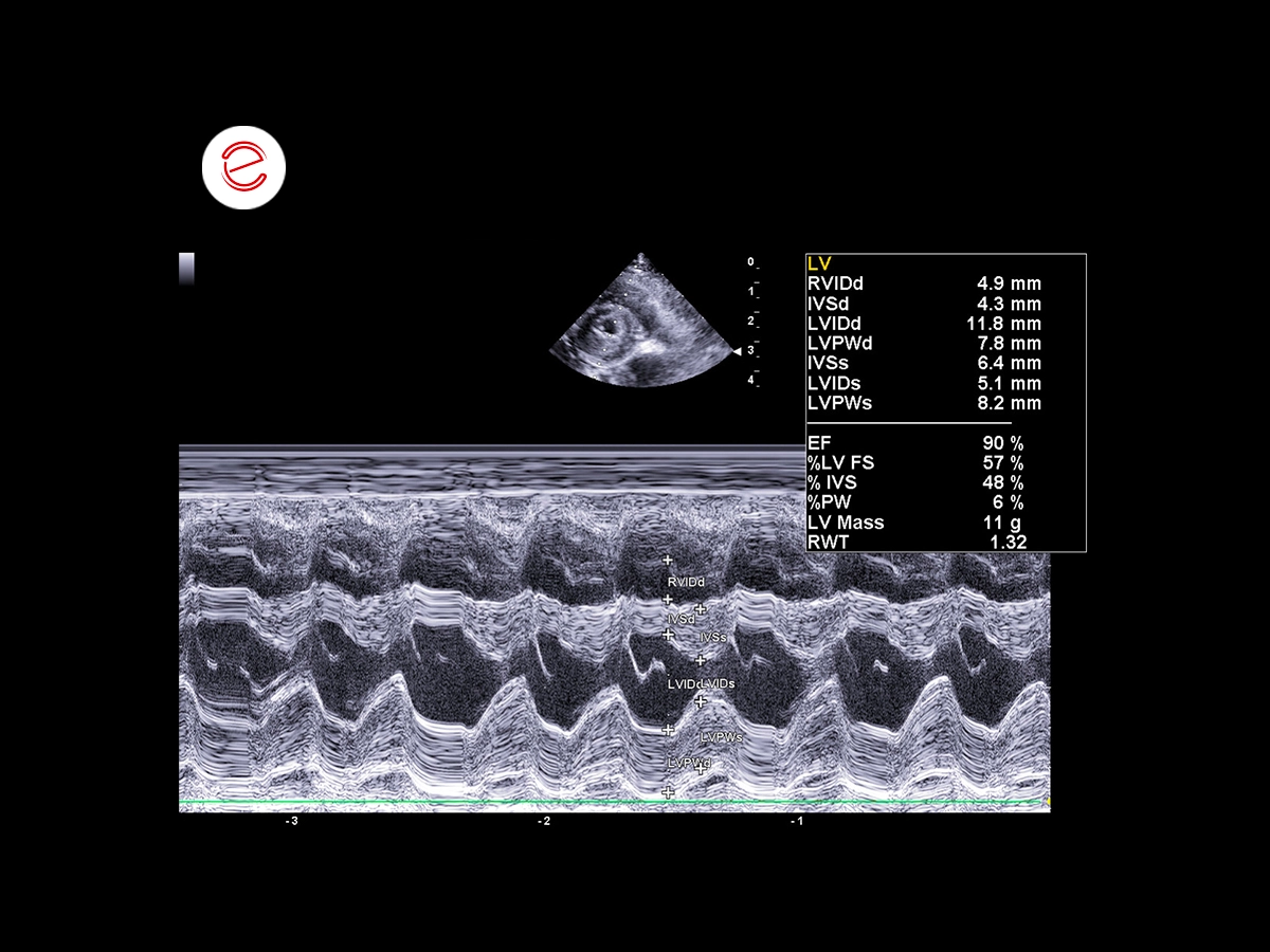
Right parasternal, short axis, M-mode
Shows hypertrophy of the free wall of the left ventricle.
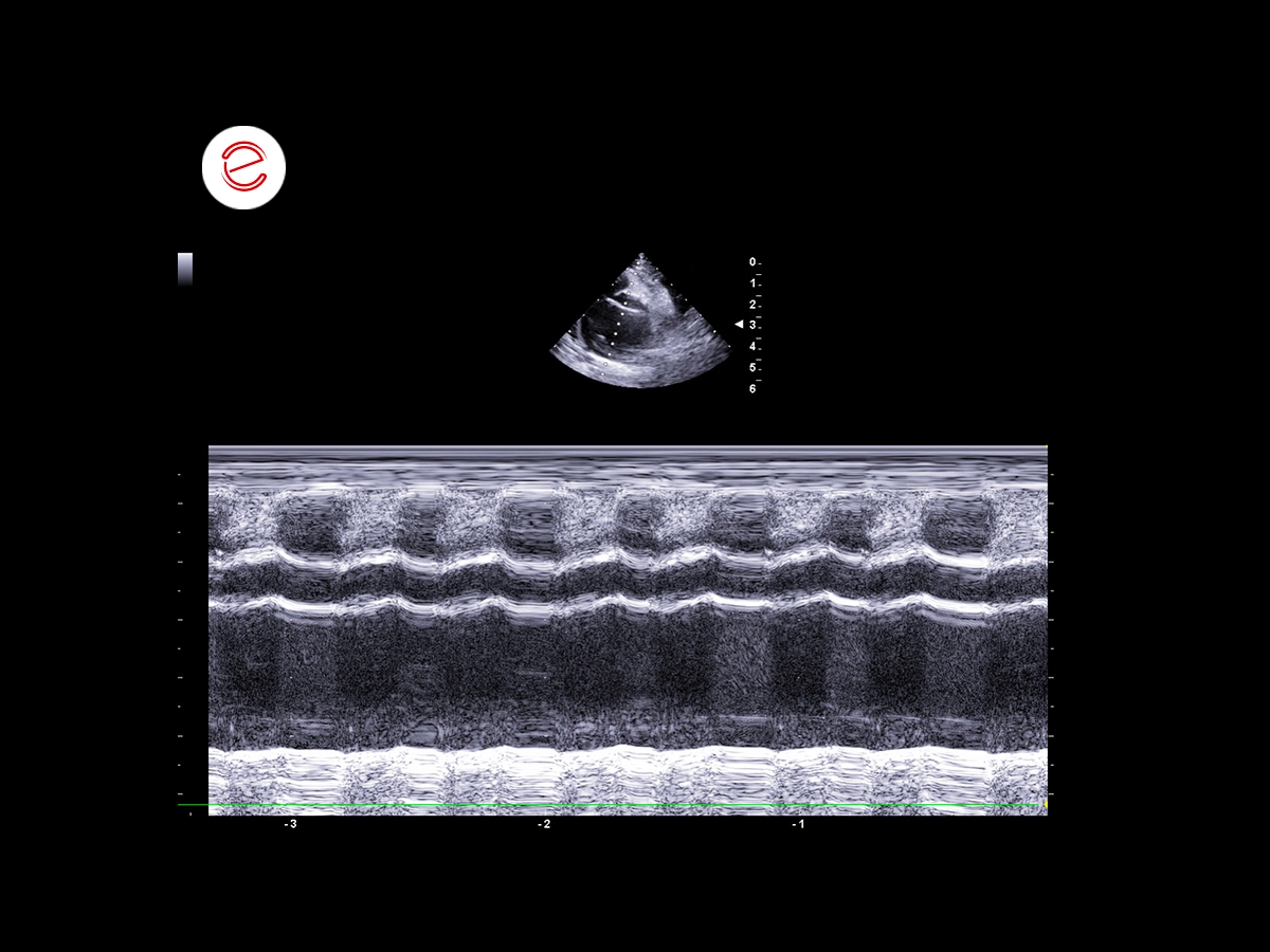
Right parasternal, short axis, Asx: Ao in M-mode
Severe left atrial enlargement with reduced kinetic energy of the atrium.
Images were acquired with the MyLab™X8VET system.
Conclusions and treatment
The complete echocardiogram examination gives a picture of restrictive cardiomyopathy, in differential diagnosis with end-stage hypertrophic cardiomyopathy, non-classified feline cardiomyopathy.
The rounded hyperechogenic structure detected inside the left auricle is presumably a thrombus.
Irregularly irregular rhythm compatible with atrial fibrillation to be confirmed by electrocardiogram trace.
Prescribed treatment: furosemide 2 mg/kg every 12 hours, clopidogrel 18.75 mg every 24 hours.
Cecilia Rossi, DVM
Clinica Veterinaria Gran Sasso, Milan, Italy.
MyLab is a trademark of Esaote spa.
Technology and features are device/configuration-dependent. Specifications subject to change without notice. Information might refer to products or modalities not yet approved in all countries. Product images are for illustrative purposes only. For further details, please contact your Esaote sales representative.
Other feline clinical cases you may be interested in
Explore the unique challenges encountered, the diagnostic procedures conducted, the solutions implemented, and the recommended treatments.