Neoplastic infiltrative process
Identification and history
- Name: Chira
- Report and medical history: Spayed female cat, 11 years old.
Patient brought in due to vomiting, loss of appetite and weight loss. On palpation of the abdomen, the presence of a neoformation was suspected and an abdominal ultrasound was therefore requested.
Diagnostics

Bladder with walls in the normal range of thickness and stratigraphy; normal anechoic endoluminal content.
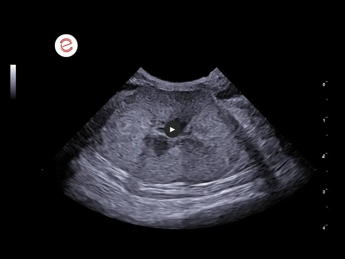
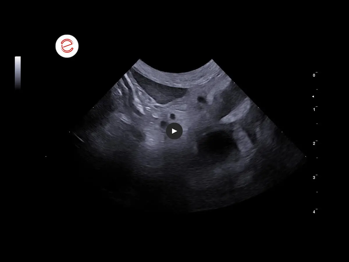
Left kidney: normal size, irregular margins, cortical hyperechogenicity, loss of cortico-medullary distinction. In the pyelic recesses, various hyperechoic areas were shown, forming a posterior shadow cone.
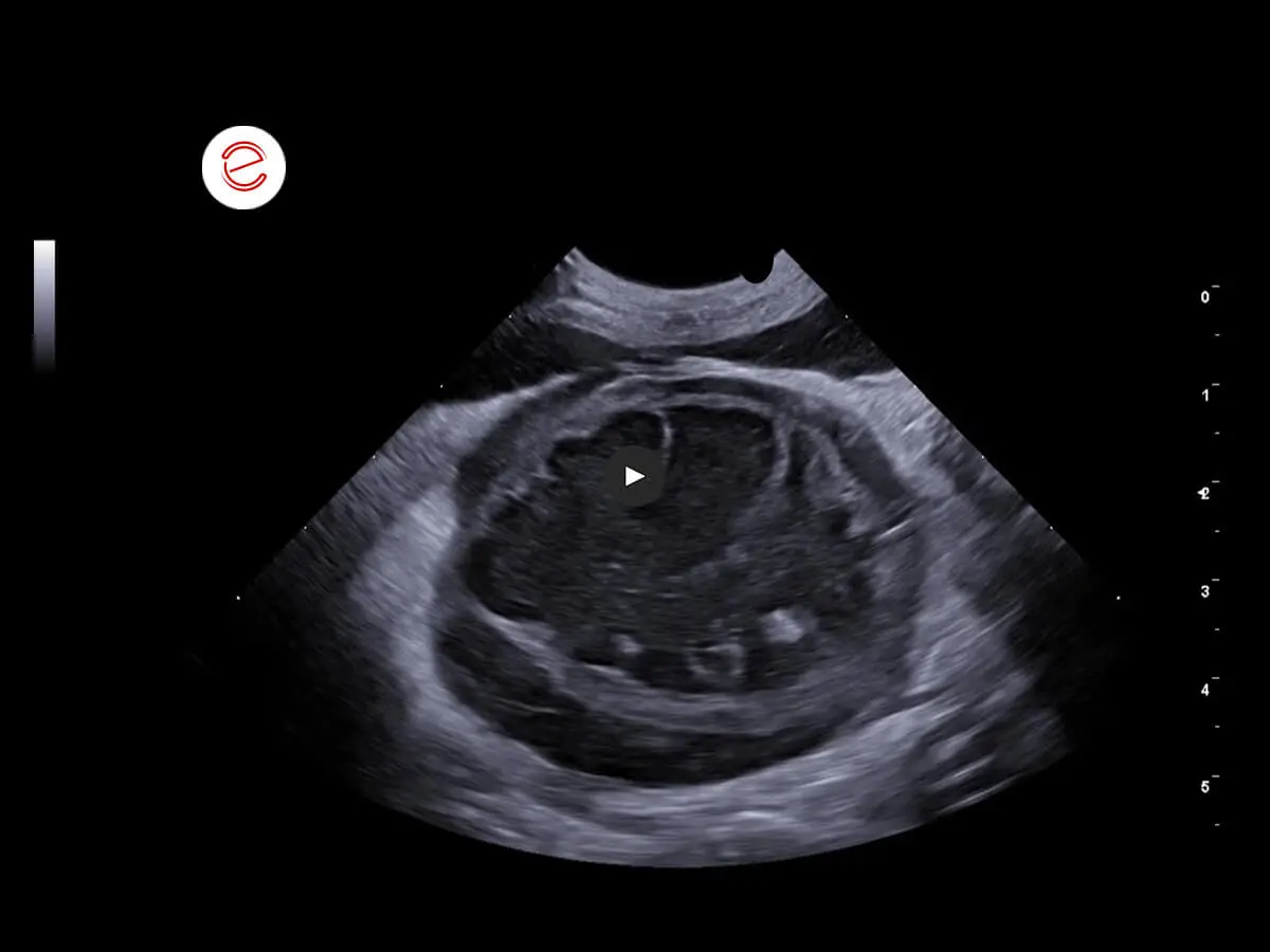
Right kidney: dimensions increased. Margins normal. No cortico-medullary distinction. Diffuse hypo-anechogenicity with findings of a hyperechoic band in which radially developing hyperechoic areas were reported.
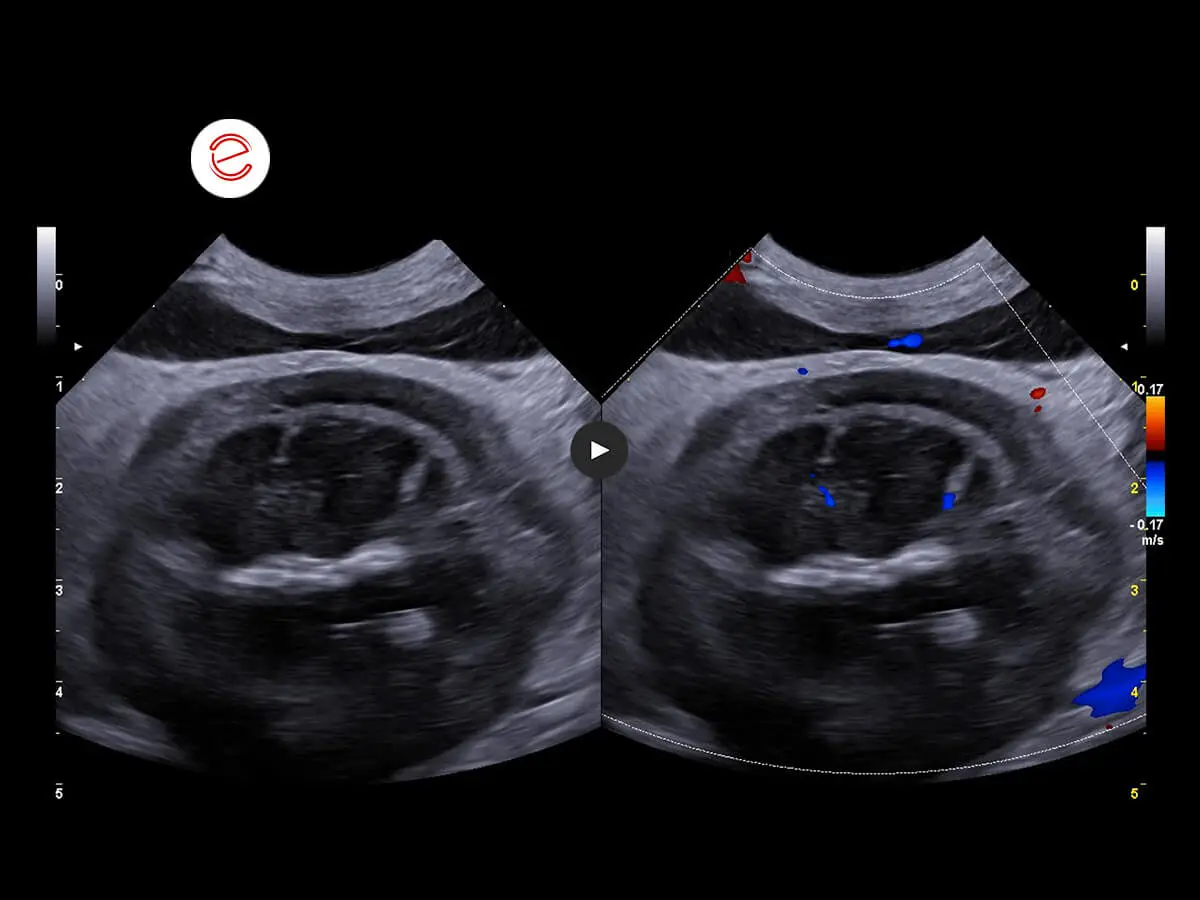
Vascularization of the right kidney preserved.
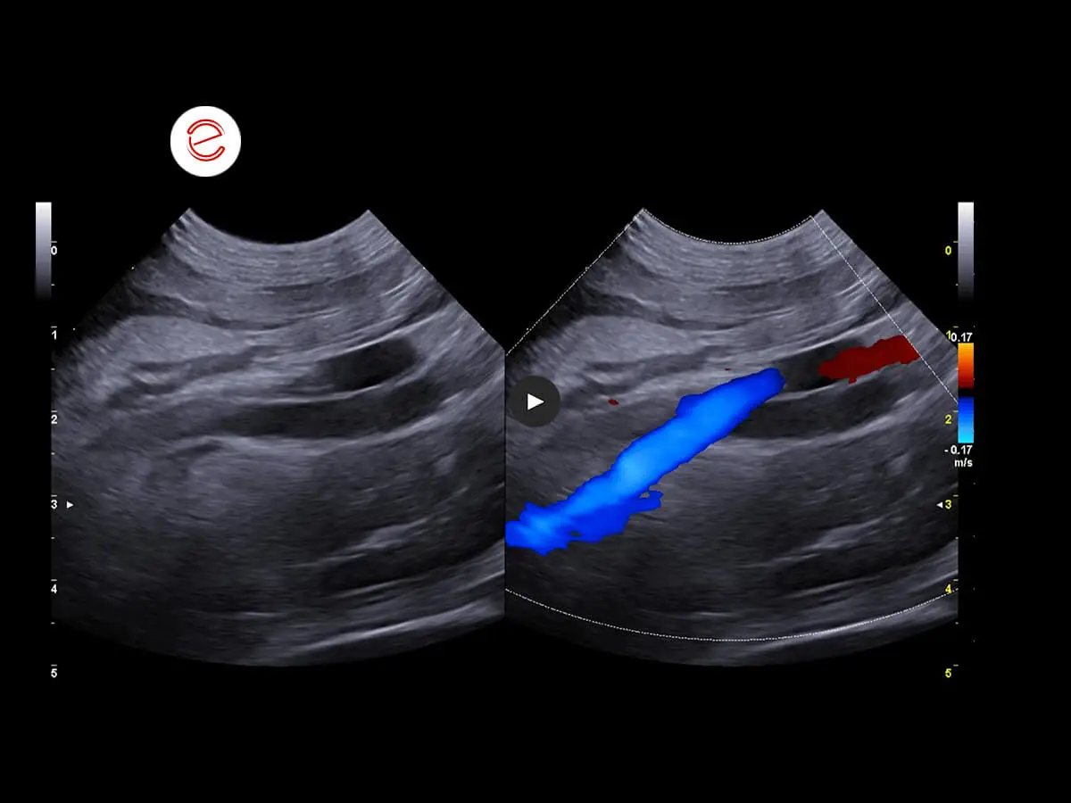
Course of the right ureter in relation to the vessels. Ureteral ectasia observed throughout its course with diffuse wall thickening.
Diffuse omental hyperechogenicity associated with findings of layers of abdominal effusion with an anechoic appearance.
Images were acquired using the MyLab™X90VET system.
Conclusions and treatment
The alterations in the right kidney are compatible with a neoplastic infiltrative process in a differential diagnosis with hydronephrosis secondary to a severe inflammatory process. Alterations in the left kidney consistent with severe nephropathy associated with findings of calcifications in differential diagnosis with lithiasis. Diffuse peritonitis also observed. Cytological examination of the right kidney recommended.
Elisabetta Boz, DVM
Clinica Veterinaria Gran Sasso, Milan, Italy

MyLab is a trademark of Esaote spa.
Technology and features are device/configuration-dependent. Specifications subject to change without notice. Information might refer to products or modalities not yet approved in all countries. Product images are for illustrative purposes only. For further details, please contact your Esaote sales representative.
Other feline clinical cases you may be interested in
Explore the unique challenges encountered, the diagnostic procedures conducted, the solutions implemented, and the recommended treatments.