Ileocolic intestinal intussusception
Identification and history
- Name: Cloe
- Report and medical history: cat, Common European, spayed female, 16 years old.
The patient presented at our clinic with dejection, anorexia, hematochezia and vomiting. On clinical examination there was depressed sensory status, BCS 3/9, 7% dehydration, and pale pink mucous membranes. Lymph nodes and respiratory rate were normal, as was the auscultation finding. On palpation of the abdomen, probable thickening of an intestinal loop was perceived.
Diagnostics
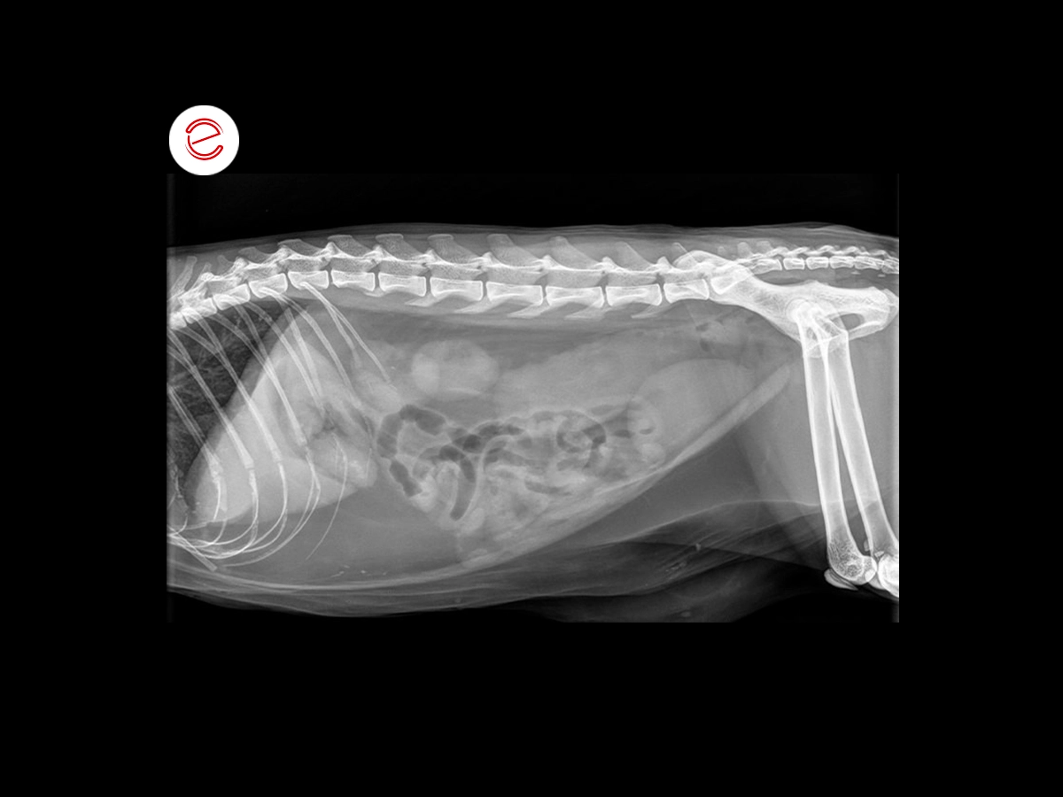
An abdominal X-ray and hematochemical tests were performed, in which an increase in creatinemia and uremia, moderate anemia with leukocytosis and neutrophilia were found. On radiography, increased radiopacity in the descending colon was found, with mild distension of the loops of the small intestine with gaseous content.
Next, an abdominal ultrasound with microconvex (Freq 3-11 MHz) and linear (Freq 4-15 MHz) probes was performed.
The abdominal scan of the liver showed the organ’s echostructure to be normal, gallbladder with normal parietal thickness, anechogenic endoluminal content with hyperechogenic material in suspension, indicative of biliary sludge.
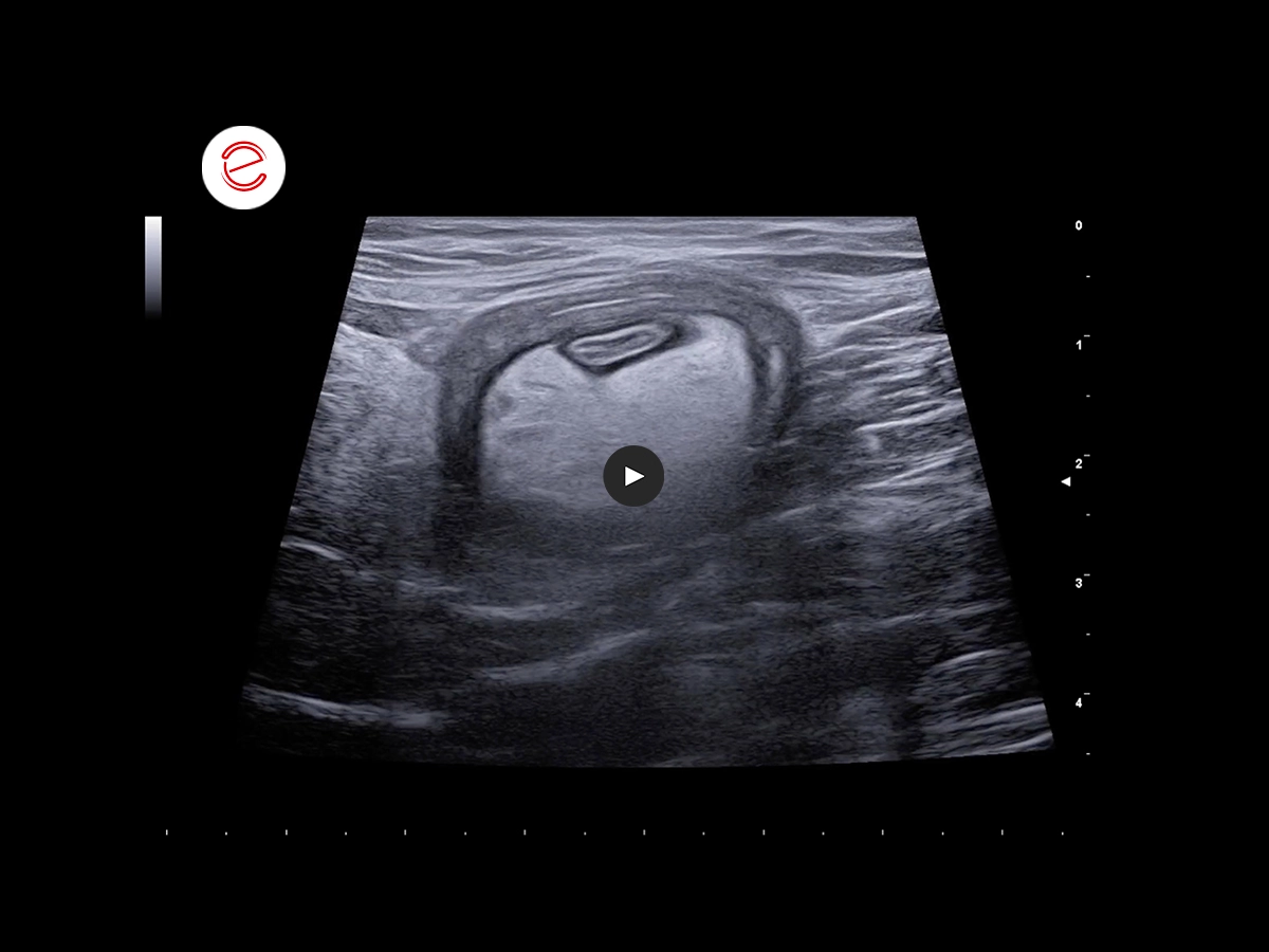
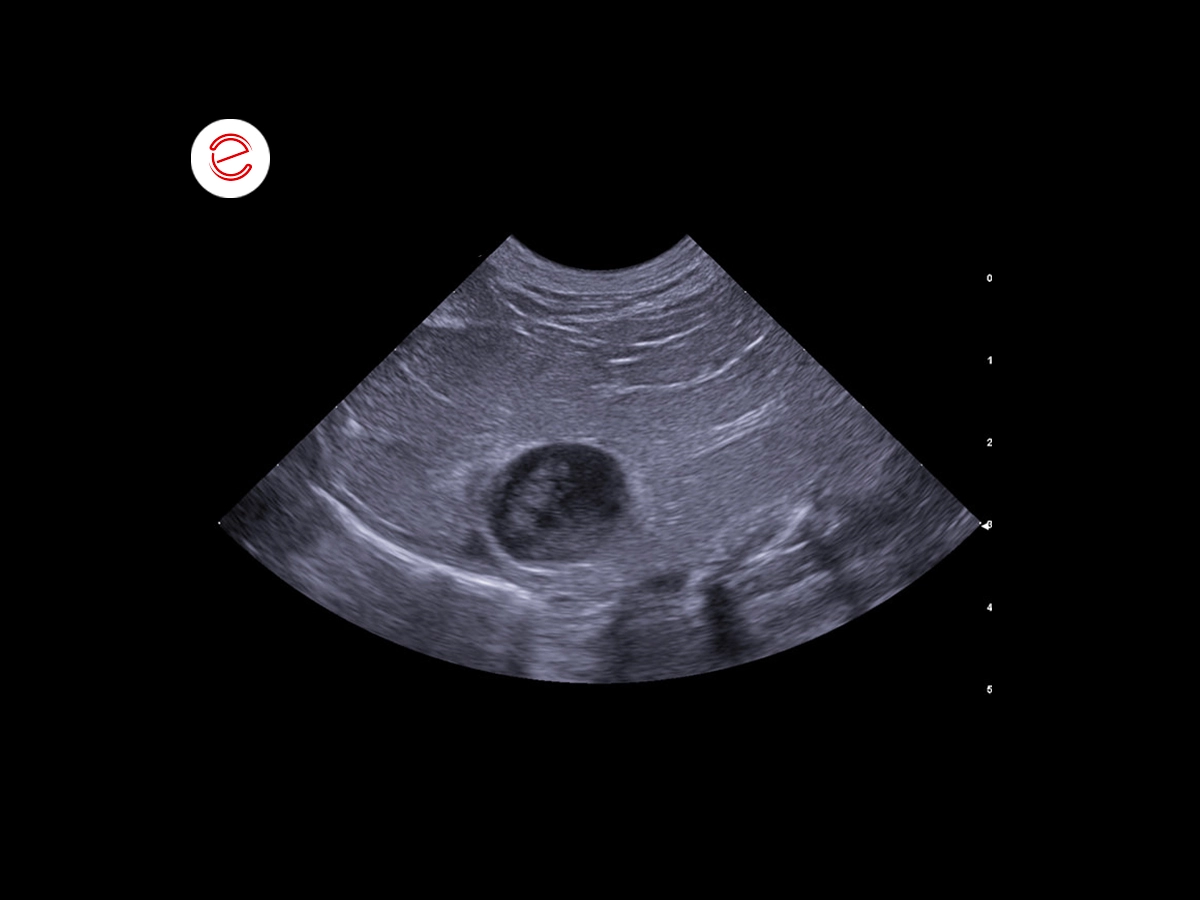
Scan of the caudal abdomen, showing a portion of double-walled intestine that assumes a concentric sign appearance in short axis. Within this portion, there is markedly hyperechogenic tissue (compatible with reactive peritoneum) and rounded hypoechogenic areas probably related to lymph nodes.
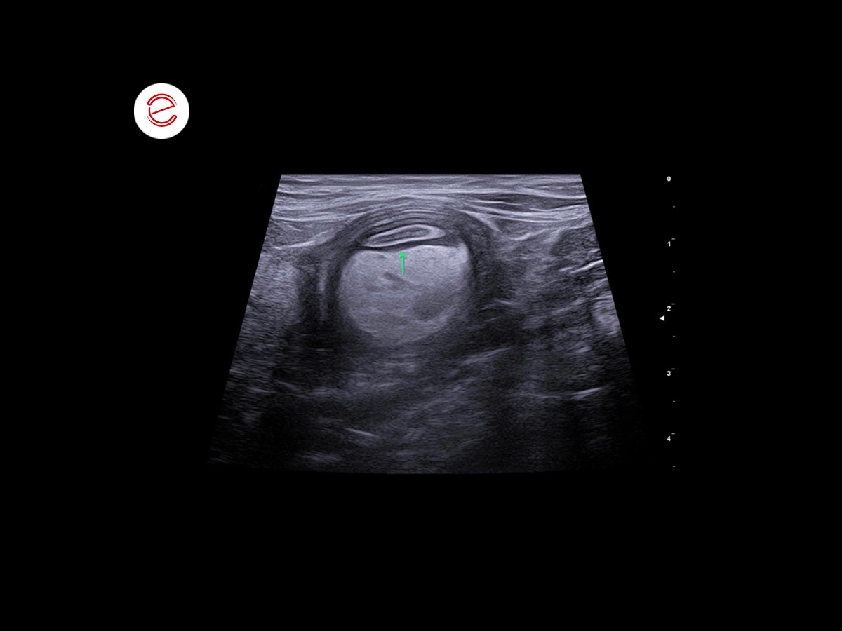
In this scan, the concentric structure is apparent, where the outer wall probably part of the colon, while the inner wall consists of small intestine (see arrow).
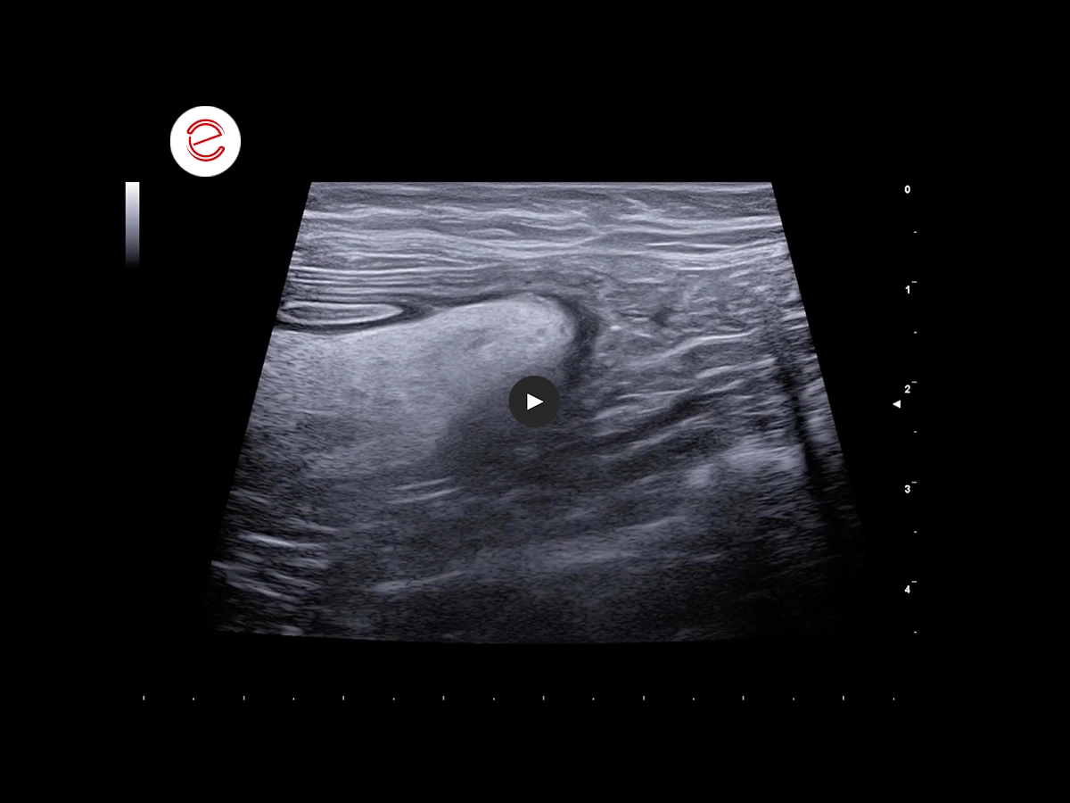
This scan confirms that the outer wall is part of the colon.
Conclusions and treatment
The finding is strongly suggestive of ileocolic intestinal intussusception. An exploratory laparotomy was performed to resolve the issue.
Small bowel enteropexy was also performed during surgery.
Melissa Papa, DVM, ECVIM-CA (Cardiology) Resident - Clinica Veterinaria Gran Sasso, Milano, Italy.

MyLab is a trademark of Esaote spa.
Technology and features are device/configuration-dependent. Specifications subject to change without notice. Information might refer to products or modalities not yet approved in all countries. Product images are for illustrative purposes only. For further details, please contact your Esaote sales representative.
Other feline clinical cases you may be interested in
Explore the unique challenges encountered, the diagnostic procedures conducted, the solutions implemented, and the recommended treatments.