Hepatic torsion
Identification and history
- Name: Felix
- Report and medical history: Rabbit, Lop-eared, M, 2 years old.
The patient presented with dragging of the hindlimb, dejection and anorexia.
Diagnostics
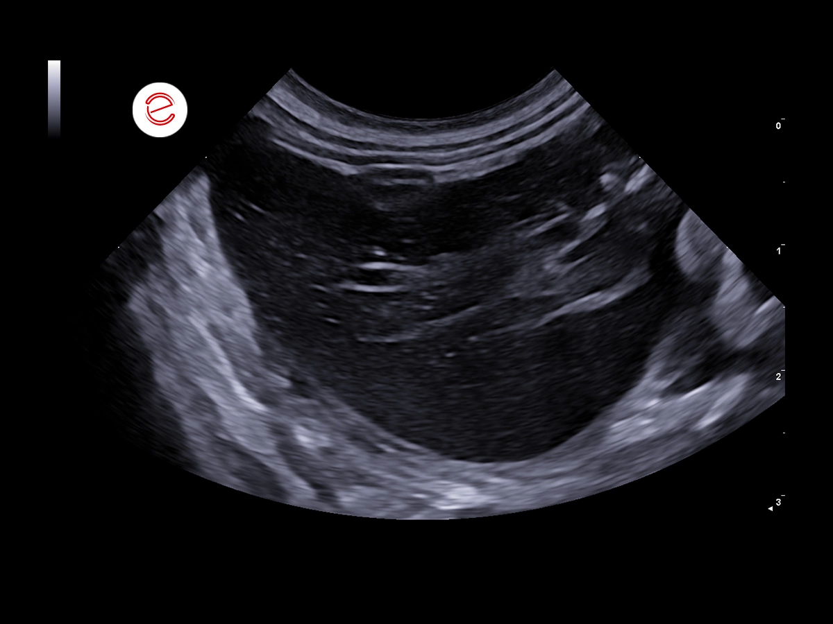
The hepatic caudate lobe was markedly increased in volume, with rounded margins, hypoechoic, heterogeneous parenchyma with a partial 'lace' pattern.
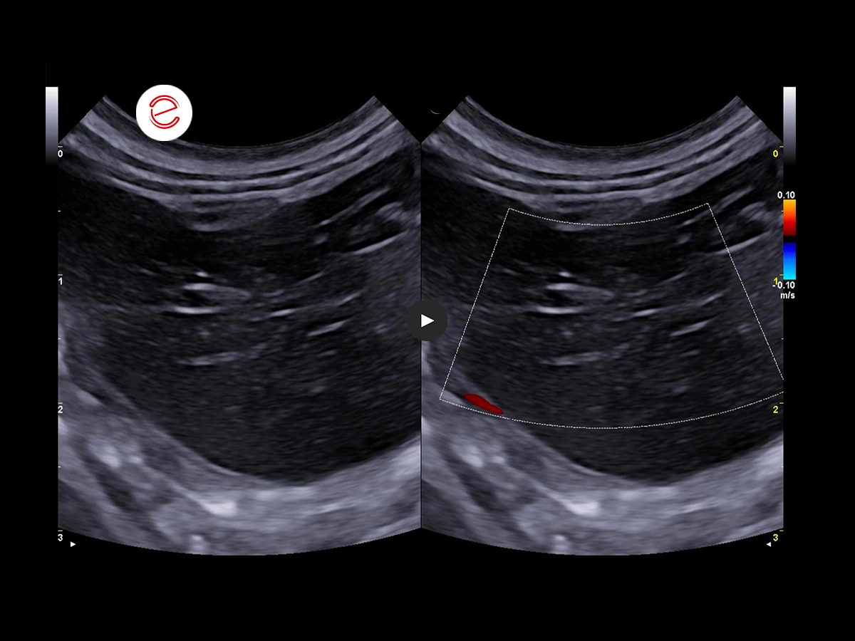
Almost complete absence of signal on Color Doppler.
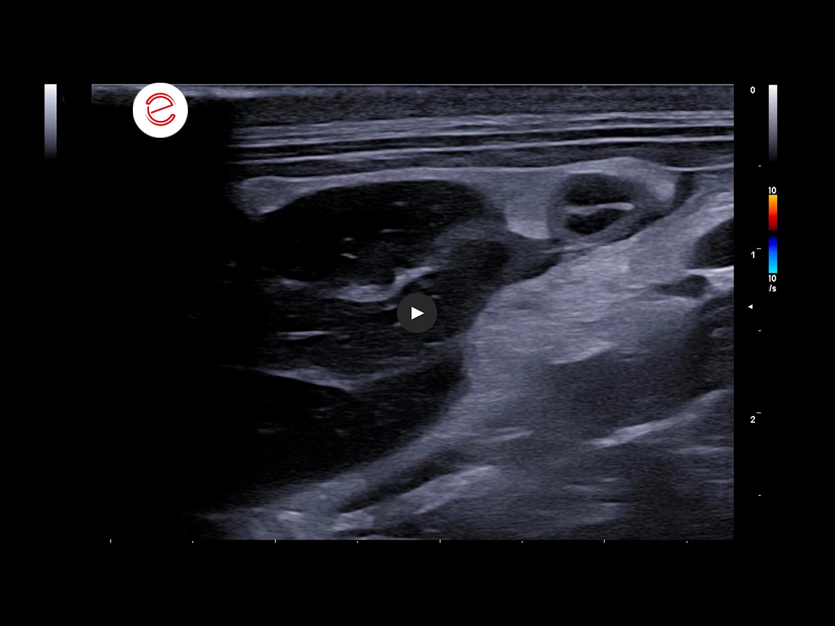
A 'whirlpool' sign was also observed in the hilar area at the level of this lobule with involvement, collapse and torsion of the portal vein of the caudate with the presence of filling defects in the hilar and parenchymal area.
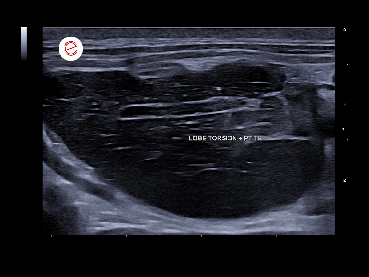
Associated thrombosis of the portal vessel within the twisted lobe.
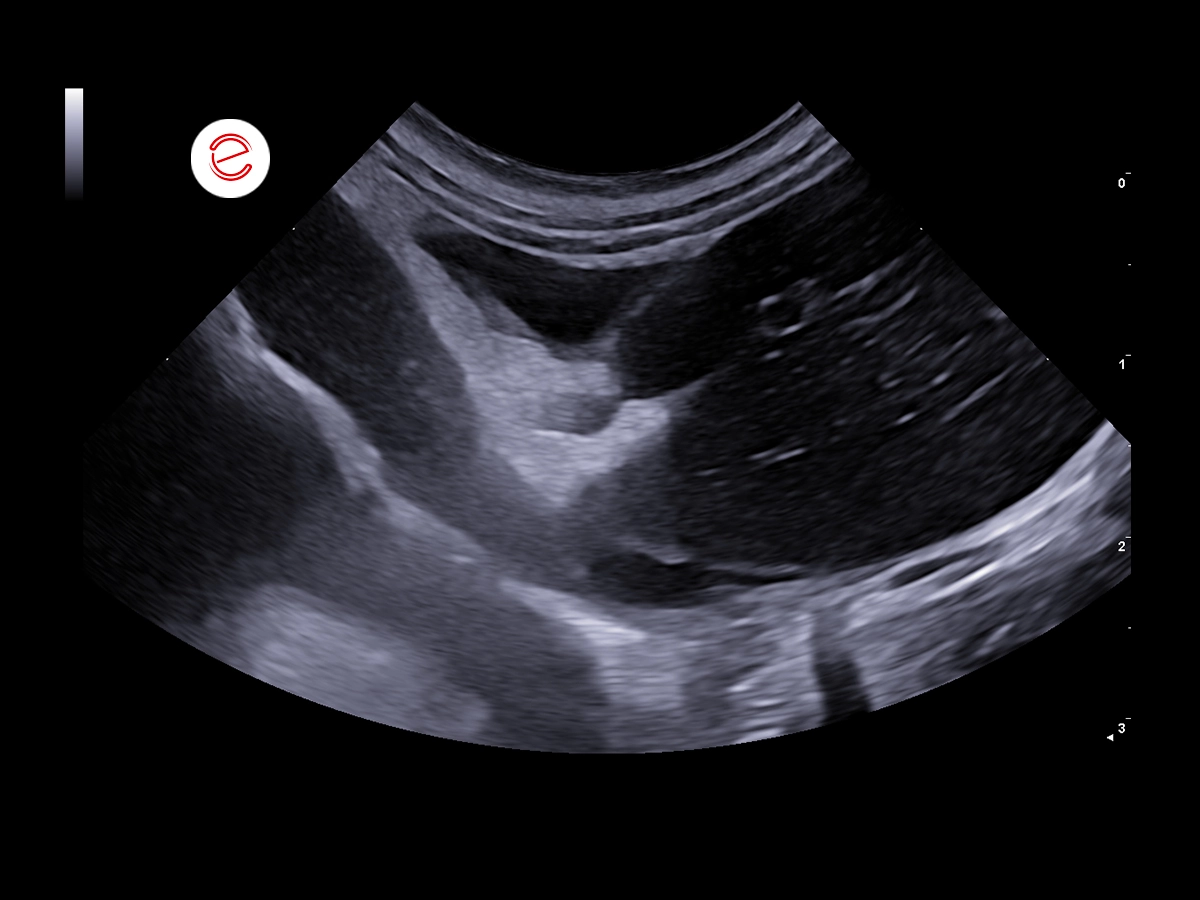
Moderate reactivity of perihepatic mesenteric adipose tissue with slight accumulation of anechoic effusion.

Pancreas moderately increased in volume with 'tiger-stripe' pattern.
Images were acquired using the MyLab™X90VET system.
Conclusions and treatment
Hepatic caudate lobe torsion with relevant portal vein thromboembolism, secondary pancreatic edema, peritoneal steatitis, and abdominal effusion (e.g. transudate vs. exudate/hemoperitoneum). Lobectomy was agreed.
Lobectomy Operation
The patient was narcotized with dexmedetomidine, ketamine, methadone; he was intubated and maintained on isoflurane. A medial incision was made starting from the xiphoid region for about 5 cm in a caudal direction with a cold scalpel blade (blade 21), the muscular planes were exposed and the linea alba and peritoneal membrane were incised in order to expose the abdominal organs. The caudate lobe was everted and appeared twisted, increased in volume, and with degenerative signs. Two knots were applied to the base of the lobe with Vycril 3/0 braided suture thread, then the lobe was removed using forceps. A square hemostatic gauze of about 1 cm on each side was applied to the stump. The remaining hepatic and abdominal portion (compatible with the topographic region of the breach) that did not show evident alterations were assessed. Sutures were applied to the muscle, subcutaneous and skin with continuous simple suture with absorbable 3/0 monofilament thread. No complications or alterations during surgery. At the end of the procedure, the patient's temperature was 37.9°C, antagonized with antipamezole IM.
University Veterinary Hospital, Department of Diagnostic Imaging, University of Milan, Lodi.
MyLab is a trademark of Esaote spa.
Technology and features are device/configuration-dependent. Specifications subject to change without notice. Information might refer to products or modalities not yet approved in all countries. Product images are for illustrative purposes only. For further details, please contact your Esaote sales representative.
Other exotics clinical cases you may be interested in
Discover the challenges faced, the examinations performed, the solutions adopted, and the treatments recommended.